

Central fibrous area in the glomerular vascular pole consists of fibrous collagens and is associated with advanced age: a cross-sectional study
- PMID: 35690711
- PMCID: PMC9188109
- DOI: 10.1186/s12882-022-02835-2
Central fibrous area in the glomerular vascular pole consists of fibrous collagens and is associated with advanced age: a cross-sectional study
Abstract
Background: For the optimal management of patients with both allograft kidneys and native kidney diseases, the recognition of the histological features associated with older age is important. This is because most pathological findings are non-specific. Central fibrous areas (CFAs) have recently been proposed to be age-related. However, the components of CFAs and whether CFAs are observed in various kidney diseases remain undetermined. This cross-sectional study was undertaken to clarify the histological features, epidemiology, and clinicopathological features of CFAs.
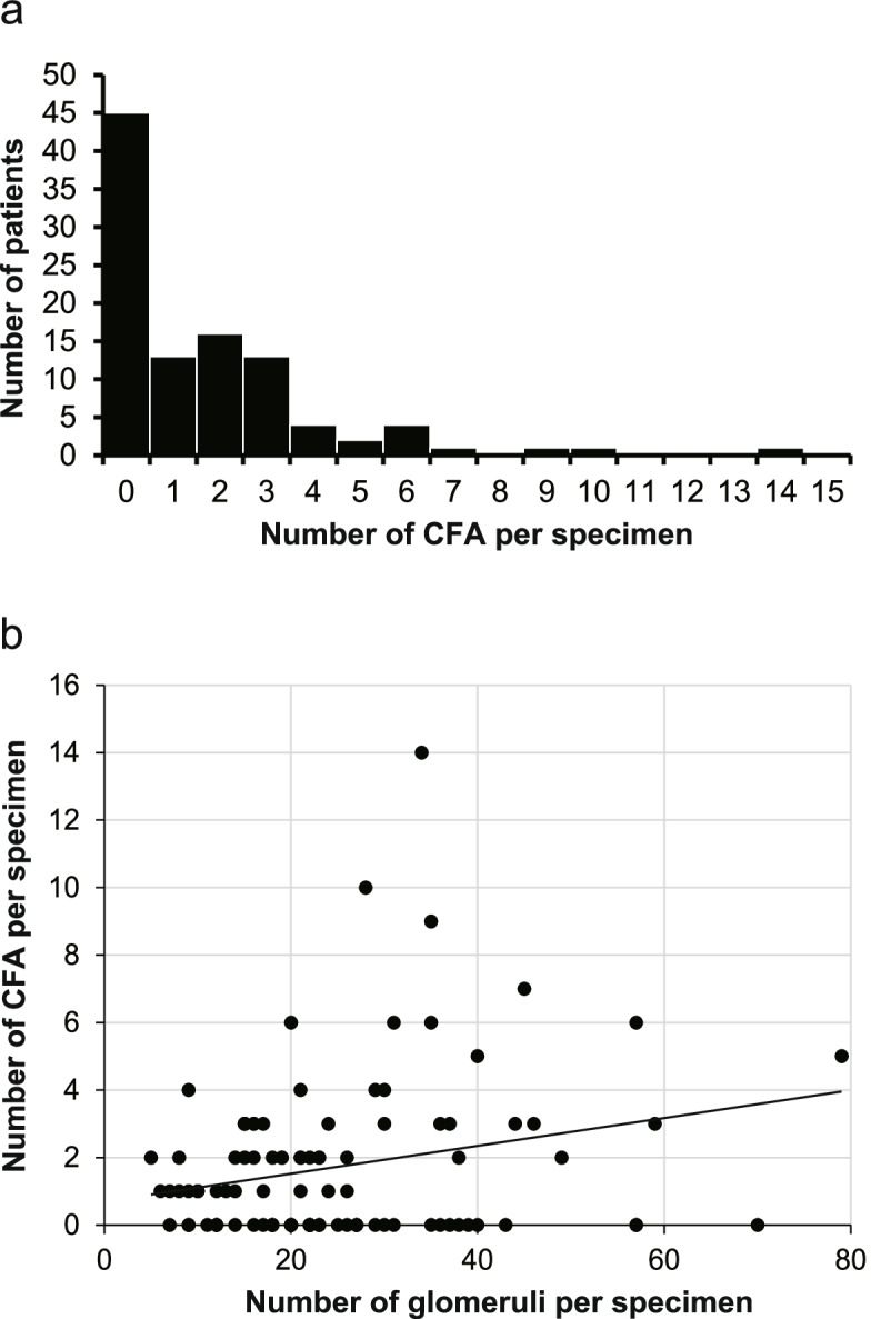
Methods: One hundred and one consecutive kidney needle biopsy specimens were retrospectively collected from seven facilities in the Hokuriku region and diagnosed at the Kanazawa University Hospital in 2015. First, the components of CFAs were analyzed using normal histostaining, immunostaining, and electron microscopy. Second, the patients were divided into two groups (CFA [+] or CFA [-]) according to the presence of CFA in the obtained samples. Clinical and histological features were compared between the two groups, and factors associated with CFA formation were determined using univariate and multivariate analyses. The number of CFAs per specimen was counted in the CFA (+) group. Third, the presence of myofibroblasts in CFA was examined by immunostaining.
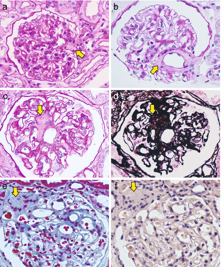
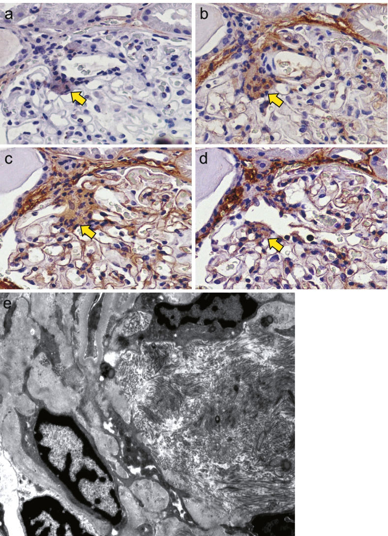
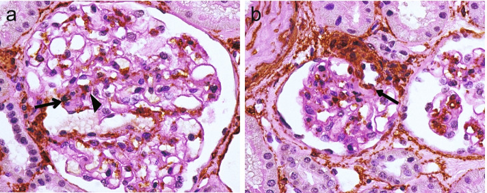
Results: CFAs were observed in 56 of 101 patients (55.4%) with various kidney diseases. CFAs consist of fibrillar collagens (collagen I and III) in addition to non-fibrillar collagens (collagen IV and VI), as confirmed by electron microscopy. Clinically, the CFA (+) group was older and had a significantly higher prevalence of hypertension and hyperlipidemia than the CFA (-) group. Histologically, elastofibrosis of the interlobular artery, arteriolar hyalinosis, and membranous nephropathy were significantly more evident in the CFA (+) group than in the CFA (-) group. Multivariate analysis revealed that older age was the sole factor associated with CFA formation. Finally, 27 of 58 (46.6%) CFA-containing glomeruli in 26 cases included alpha-smooth muscle actin-positive cells in or adjacent to the CFA.
Conclusions: CFAs consist of fibrous collagens in addition to matrix collagens. CFA formation is associated with older age and was observed in various kidney diseases.
Keywords: Central fibrous area; Collagen; Myofibroblast; Older age.
© 2022. The Author(s).
Conflict of interest statement
The authors declare that they have no competing interests.
Figures
Similar articles
-
Central fibrous areas: changes in glomerular vascular pole lesions associated with age and disease.Int Urol Nephrol. 2022 Sep;54(9):2263-2273. doi: 10.1007/s11255-022-03126-3. Epub 2022 Jan 31. Int Urol Nephrol. 2022. PMID: 35099688 Free PMC article.
-
Age-related nephropathy in the Fischer 344 rat is associated with overexpression of collagens and collagen-binding heat shock protein 47.Cell Tissue Res. 1998 Sep;293(3):471-8. doi: 10.1007/s004410051139. Cell Tissue Res. 1998. PMID: 9716737
-
Stem cell factor and crescentic glomerulonephritis.Am J Kidney Dis. 2003 Apr;41(4):785-95. doi: 10.1016/s0272-6386(03)00026-x. Am J Kidney Dis. 2003. PMID: 12666065
-
Cyclopropane ring formation in membrane lipids of bacteria.Microbiol Mol Biol Rev. 1997 Dec;61(4):429-41. doi: 10.1128/mmbr.61.4.429-441.1997. Microbiol Mol Biol Rev. 1997. PMID: 9409147 Free PMC article. Review.
-
Non-fibrillar collagens: key mediators of post-infarction cardiac remodeling?J Mol Cell Cardiol. 2010 Mar;48(3):530-7. doi: 10.1016/j.yjmcc.2009.06.017. Epub 2009 Jun 30. J Mol Cell Cardiol. 2010. PMID: 19573533 Review.
References
MeSH terms
Substances
LinkOut - more resources
Full Text Sources
Medical
