. 2022 Jun 11;12(1):86.
doi: 10.1186/s13578-022-00809-3.

Inclusion of a dual signal sequence enhances the immunogenicity of a novel viral vectored vaccine against the capsular group B meningococcus

Affiliations

Inclusion of a dual signal sequence enhances the immunogenicity of a novel viral vectored vaccine against the capsular group B meningococcus

Dylan Sheerin et al. Cell Biosci. .

Abstract

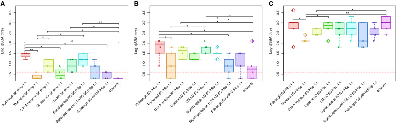
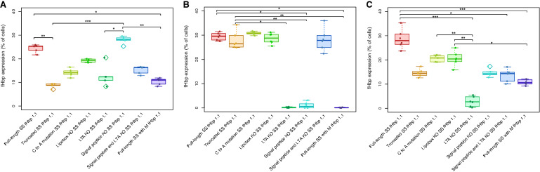
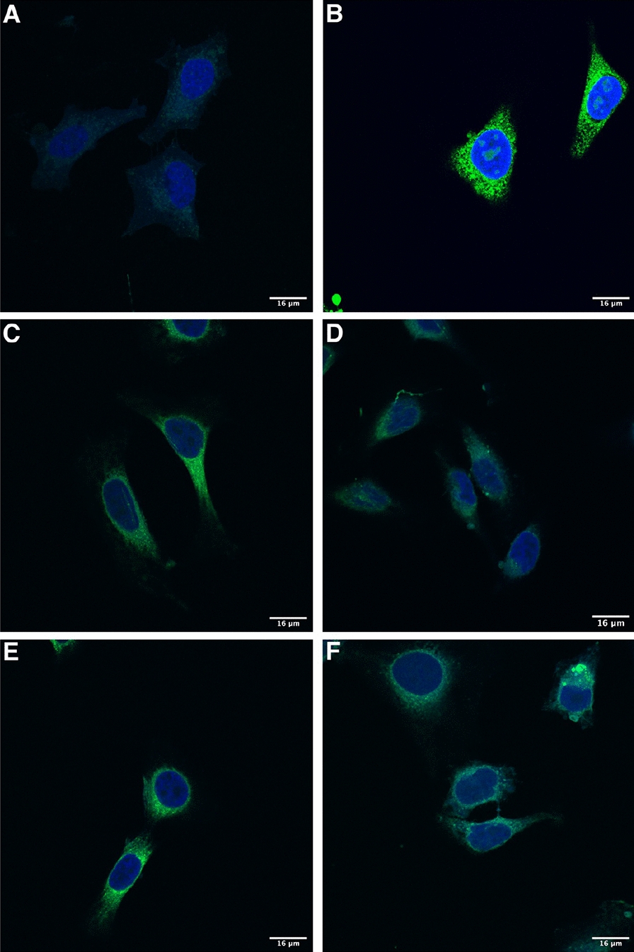
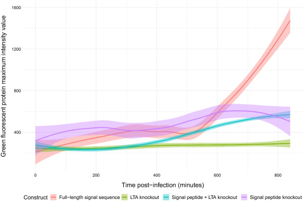
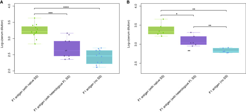
Background: Disease caused by the capsular group B meningococcus (MenB) is the leading cause of infectious death in UK infants. A novel adenovirus-based vaccine encoding the MenB factor H binding protein (fHbp) with an N-terminal dual signal sequence induces high titres of protective antibody after a single dose in mice. A panel of N-terminal signal sequence variants were created to assess the contribution of components of this sequence to transgene expression kinetics of the encoded antigen from mammalian cells and the resultant effect on immunogenicity of fHbp.

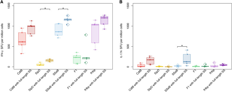
Results: The full-length signal sequence (FL SS) resulted in superior early antigen expression compared with the panel of variants, as measured by flow cytometry and confocal imaging, and supported higher bactericidal antibody levels against the expressed antigen in mouse sera < 6 weeks post-immunisation than the licensed four component MenB vaccine. The FL SS also significantly increased antigen-specific T cell responses against other adenovirus-encoded bacterial antigens in mice.

Conclusions: These findings demonstrate that the FL SS enhances immunogenicity of the encoded antigen, supporting its inclusion in other viral vectored bacterial antigen transgenes.

Keywords: Expression kinetics; Meningococcal disease; Signal sequence; Transgene; Viral vector vaccines.


Conflict of interest statement

A.J.P. is Chair of UK Dept. Health and Social Care’s (DHSC) Joint Committee on Vaccination & Immunisation (JCVI) and is a member of the WHO’s SAGE. The views expressed in this article do not necessarily represent the views of DHSC, JCVI, NIHR or WHO. The University of Oxford has entered into a partnership with AstraZeneca on coronavirus vaccine development. C.S.R., C.D., D.S., and A.J.P. are named inventors on a patent application in the field of meningococcal vaccine. A.J.P waives all his rights to any patent.

Figures

Fig. 1
Fig. 1
Serum bactericidal antibody (SBA) titres in sera of mice immunised with human adenovirus encoding N-terminal signal sequence variants of the factor H binding protein. Groups of six BALB/c mice were immunised with a sub-optimal dose of 1 × 107 infectious units of one of the signal sequence variant constructs or 1/10 of the human dose of 4CMenB (two-dose regimen administered at day 0 and day 21) and SBA assays were performed against the H44/76 strain of Neisseria meningitidis using sera derived from blood samples taken at different timepoints post-immunisation. A Week two SBA titres. B Week four SBA titres. C Week six SBA titres. The dotted red line represents the cut-off titre of 1:4 deemed sufficient for protection. Differences in the SBA titre between constructs are attributable to the N-terminal signal sequence variants of the factor H binding protein, particularly at early timepoints post-immunisation. Statistical comparisons were made using a Mann–Whitney U-test. *p < 0.05; **p < 0.01
Fig. 2
Fig. 2
Expression of human adenovirus-encoded factor H binding protein N-terminal signal sequence variants from HeLa cells. A HeLa cells (1 × 106 per sample) were infected overnight with 5 × 108 infectious units of one of a series of human adenovirus serotype 5 (AdHu5) constructs encoding an N-terminal sequence variant of the factor H binding protein (fHbp) and expression was quantified by flow cytometry after surface and intracellular staining of harvested cells with an anti-fHbp antibody (JAR5) and a fluorescently-tagged detection antibody. Cells were tested for intracellular expression by stimulation with brefeldin A to stop protein transport within the cells and harvested at B three-hour and C five-hour timepoints post-infection to measure early expression levels by intracellular staining. The y-axis corresponds to the percentage of total fluorescent (fHbp-expressing) HeLa cells after overnight infection. The amino acid composition of the N-terminal sequence impacts upon the expression of the transgene-encoded antigen within the first 24 h of infection. Statistical comparisons were made using a Mann–Whitney U-test. *p < 0.05; **p < 0.01; ***p < 0.001
Fig. 3
Fig. 3
Confocal images of HeLa cells infected overnight human adenovirus encoding factor H binding protein (fHbp) N-terminal signal sequence (SS) variants. N-terminal signal sequence variant transgenes were designed to include an enhanced green fluorescent protein (eGFP) sequence and cloned into adenovirus vectors. Representative images for A Uninfected cells (negative control), B eGFP only (positive control), C Full-length SS fHbp 1.1-eGFP, D LTA knockout (KO) SS fHbp 1.1-eGFP, E Signal peptide (SP) KO SS fHbp 1.1-eGFP and F SP + LTA KO SS fHbp 1.1-eGFP. Blue fluorescence indicates DAPI-stained nuclei, green fluorescence indicates antigen-eGFP expression. 16 μm scale bars are shown in the bottom right corner of each image
Fig. 4
Fig. 4
Expression of factor H binding protein N-terminal signal sequence variants from HeLa cells infected with adenovirus vectors over the course of 14 h. N-terminal signal sequence variant transgenes were designed to include an enhanced green fluorescent protein sequence and cloned into adenovirus vectors. Green fluorescent protein intensity values were calculated from images taken across three separate fields of the infected wells and averaged for each signal sequence variant. Loess smoothing was applied to trend lines for each construct and 95% confidence intervals are shaded
Fig. 5
Fig. 5
Anti-F1 antigen IgG titres in mouse sera post-immunisation with human adenovirus vectors encoding signal sequence variants of the F1 antigen. Groups of 12 BALB/c mice were immunised with a sub-optimal dose of 1 × 107 infectious units of one of the human adenovirus serotype 5 (AdHu5) vectors encoding the Yersinia pestis F1 antigen with native signal sequence (SS), a heterologous N-terminal full-length (FL) SS, or a truncated form of the F1 antigen lacking any SS. Enzyme-linked immunosorbent assays were performed on serum samples taken at weeks A two and B four post-immunisation to determine the titres of anti-F1 antigen IgG in sera. The humoral response induced by the F1 antigen with native SS was superior to that induced by the incorporation of the heterologous FL SS to this antigen or its truncated form at both timepoints. Statistical comparisons were made using a Mann–Whitney U-test. *p < 0.05; **p < 0.01; ***p < 0.001; ****p < 0.0001
Fig. 6
Fig. 6
Antigen-specific T cell responses induced in mice two weeks after immunisation with human adenovirus vectors encoding bacterial antigens with or without an N-terminal signal sequence. Groups of six BALB/c mice were immunised with human adenovirus serotype 5 (AdHu5) vaccines expressing one of a series of bacterial antigens, with or without an N-terminal signal sequence (SS). Spleens were harvested two weeks post-immunisation, processed, and stimulated at a concentration of 3 μg/mL with the relevant peptide pool. An interferon (IFN)-γ/interleukin (IL)-17A dual colour fluorospot was performed to assess antigen-specific T cell responses associated with these cytokines. A IFN-γ and B IL-17A spot-forming units (SFU) per million cells were quantified for each antigen. Inclusion of an N-terminal SS was found to boost both types of antigen-specific T cell responses. Statistical comparisons were made using a Mann–Whitney U-test. *p < 0.05

Similar articles

Cited by

References

    1. Finne J, et al. An IgG monoclonal antibody to group B meningococci cross-reacts with developmentally regulated polysialic acid units of glycoproteins in neural and extraneural tissues. J Immunol. 1987;138(12):4402–4407. - PubMed
    1. Rollier CS, et al. The capsular group B meningococcal vaccine, 4CMenB : clinical experience and potential efficacy. Expert Opin Biol Ther. 2015;15(1):131–142. doi: 10.1517/14712598.2015.983897. - DOI - PubMed
    1. Gandhi A, Balmer P, York LJ. Characteristics of a new meningococcal serogroup B vaccine, bivalent rLP2086 (MenB-FHbp; Trumenba®) Postgrad Med. 2016;128(6):548–556. doi: 10.1080/00325481.2016.1203238. - DOI - PubMed
    1. Committee on Infectious Diseases Recommendations for Serogroup B Meningococcal Vaccine for Persons 10 Years and Older. Pediatrics. 2016 doi: 10.1542/peds.2016-1890. - DOI - PubMed
    1. Christensen H, et al. Re-evaluating cost effectiveness of universal meningitis vaccination (Bexsero) in England: modelling study. BMJ. 2014;349:g5725. doi: 10.1136/bmj.g5725. - DOI - PMC - PubMed

LinkOut - more resources