

Positive dysphotopsia after intrascleral intraocular lens fixation: a case report
- PMID: 35690806
- PMCID: PMC9188193
- DOI: 10.1186/s12886-022-02474-z
Positive dysphotopsia after intrascleral intraocular lens fixation: a case report
Abstract
Background: Positive dysphotopsia is a symptom caused by the reflection of incident light through the pupil at the inner surface of the intraocular lens (IOL) edge after cataract surgery and is perceived as an abnormal arcuate or radiating photopic image at night or indoors with a light source. Although positive dysphotopsia is one of the most important symptoms that affect patients after cataract surgery, it is still not well known even among ophthalmologists. Positive dysphotopsia as the cause of patient complaint following intraocular surgery other than cataract surgery has not been identified.
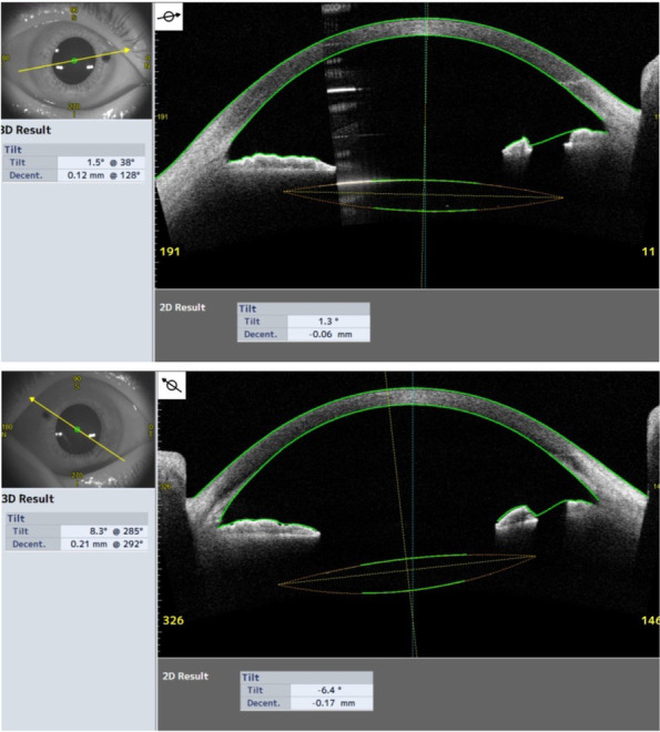
Case presentation: A 52-year-old man underwent IOL extraction and intrascleral IOL fixation for bilateral IOL subluxation at another hospital. The right eye had good subjective visibility, but the patient noticed symptoms of light sources appearing divided into multiple lights indoors after surgery in the left eye. Because the cause of the symptoms could not be identified, the patient visited our department. At the time of his first visit, the corrected visual acuity in both eyes was good, and ocular findings in eye position, motility, intraocular pressure, and fundus were within normal limits. The elongated holes of peripheral iridectomy (PI) created during previous intrascleral IOL fixation were observed to be approximately 2 mm in length on the nasal side in both eyes. The PI hole in the right eye was covered by the optics of the IOL, whereas the edge of the IOL overlapped the center of the PI hole in the left eye. Accordingly, we concluded that the abnormal photopic image in the left eye was caused by positive dysphotopsia, in which light passing through the PI hole was reflected by the edge of the IOL. We attempted surgical closure of the PI hole, resulting in the complete disappearance of positive dysphotopsia.
Conclusions: A PI hole created during intrascleral IOL fixation may cause postoperative positive dysphotopsia depending on the position of the IOL edge. Thus, surgeons should be aware of the importance of the size and location of the PI hole when creating it during surgery.
Keywords: Intraocular lens edge; Intrascleral intraocular lens fixation; Peripheral iridectomy; Positive dysphotopsia.
© 2022. The Author(s).
Conflict of interest statement
The authors declare that they have no competing interests.
Figures


References
-
- McCannel MA. A retrievable suture idea for anterior uveal problems. Ophthalmic Surg. 1976;7(2):98–103. - PubMed
Publication types
MeSH terms
LinkOut - more resources
Full Text Sources
Medical
Research Materials
Miscellaneous
