. 2022 Jun 11;19(1):18.
doi: 10.1186/s12983-022-00464-x.

Generation and characterization of genome-modified chondrocyte-like cells from the zebra finch cell line immortalized by c-MYC expression

Affiliations

Generation and characterization of genome-modified chondrocyte-like cells from the zebra finch cell line immortalized by c-MYC expression

Kyung Min Jung et al. Front Zool. .

Abstract

Background: Due to their cost effectiveness, ease of use, and unlimited supply, immortalized cell lines are used in place of primary cells for a wide range of research purposes, including gene function studies, CRISPR-based gene editing, drug metabolism tests, and vaccine or therapeutic protein production. Although immortalized cell lines have been established for a range of animal species, there is still a need to develop such cell lines for wild species. The zebra finch, which is used widely as a model species to study the neurobiological basis of human speech disorders, has been employed in several functional studies involving gene knockdown or the introduction of exogenous transgenes in vivo; however, the lack of an immortalized zebra finch cell line has hampered precise genome editing studies.

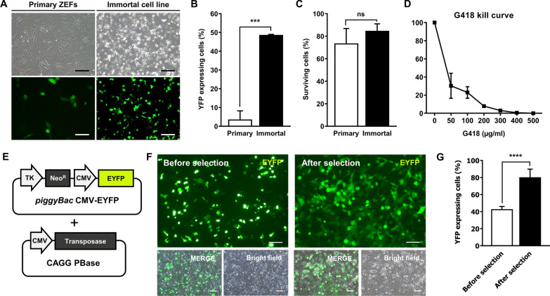
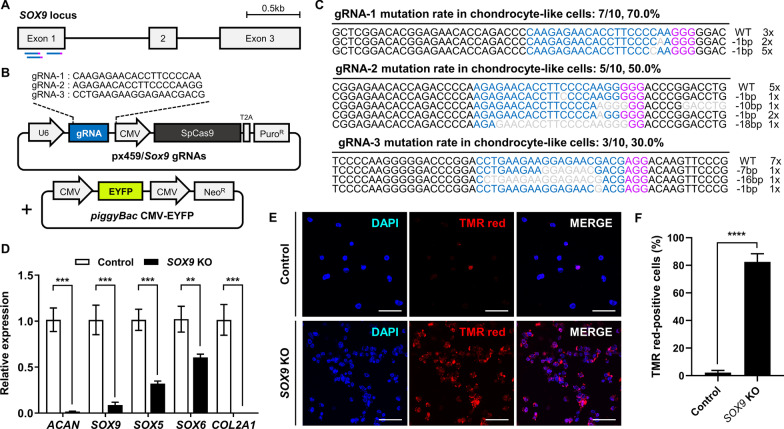
Results: Here, we established an immortalized cell line by a single genetic event, expression of the c-MYC oncogene, in zebra finch embryonic fibroblasts and examined its potential suitability for gene targeting investigations. Retroviral vector-mediated transduction of c-MYC was used to immortalize zebra finch primary fibroblasts; the transformed cells proliferated stably over several passages, resulting in the expression of chondrocyte-specific genes. The transfection efficiency of the immortalized cells was much higher than that of the primary cells. Targeted knockout of the SOX9 gene, which plays a role in the differentiation of mesenchymal progenitor cells into chondrocytes, was conducted in vitro and both apoptosis and decreased expression levels of chondrogenic marker genes were observed in edited cells.

Conclusions: The c-MYC induced immortalized chondrocyte-like cell line described here broadens the available options for establishing zebra finch cell lines, paves the way for in-depth biological researches, and provides convenient approaches for biotechnology studies, particularly genomic modification research.

Keywords: Chondrocyte-like cells; Gene editing; Immortalized cell line; Zebra finch; c-MYC.


Conflict of interest statement

The authors declare that they have no competing interests.

Figures

Fig. 1
Fig. 1
Immortalization of primary zebra finch embryonic fibroblasts (ZEFs) by c-MYC expression. A The growth curve of primary ZEFs. Subculturing was performed on days 5, 10, 15, and 20. PDL, population doubling level. B Schematic illustration of the immortalization of primary ZEFs via retroviral vector-mediated transduction. C The morphology of c-MYC-treated cells. Scale bar, 100 μm. D The growth curve of c-MYC-treated cells. E Cellular senescence of primary cells and the immortalized cell line. The black arrowhead indicates a stained (scenescent) cell. Scale bar, 100 μm. SA‐β‐Gal, senescence‐associated-β-galactosidase. F The percentages of senescent primary and immortalized cells; **p < 0.01. G Cell cycle analyses of primary cells and the immortalized cell line. H Karyotyping analysis of primary cells at passage 1 and immortalized cells at passage 65. Karyotyping analysis only involved in the macrochromosomes of zebra finch. I Telomerase activity of the immortalized cell line at passages 1, 31, and 72. The gene expression levels of ectopic mouse c-MYC from the pMXs-c-MYC plasmid and endogenous zebra finch c-MYC, TERT, and RB1 were analyzed by RT-PCR. DW, distilled water
Fig. 2
Fig. 2
Characterization of the immortalized zebra finch cell line. A Alcian blue staining of primary zebra finch embryonic fibroblasts (ZEFs) and the immortalized cell line. Scale bar, 100 μm. B Gene expression profiling of the primary ZEFs, the immortalized zebra finch cell line, and primary chondrocyte-like cells (CLCs). DW, distilled water. C Quantitative RT-PCR analyses of the primary and immortalized cells; *p < 0.05, ***p < 0.0005, ****p < 0.0001
Fig. 3
Fig. 3
Enhanced transfection efficieny of the chondrocyte-like immortalized zebra finch cell line. A Representative images of transfected immortalized cells and primary zebra finch embryonic fibroblasts (ZEFs). Scale bar, 100 μm. B Transfection efficiencies of the primary and immortalized cells, as determined by flow cytometry. C The viabilities of the transfected primary and immortalized cells, determined via manual counting after trypan blue staining; ***p < 0.0001. D The sensitivity of chondrocyte-like zebra finch immortalized cells to G418. The cells were exposed to increasing concentrations of G418 for 1 week and the percentage survival was determined by trypan blue staining. E Schematic overview of the piggyBac YFP expression plasmid and the CAGG PBase plasmid. F Images of YFP-expressing immortalized cells before and after selection with G418 (300 μg/mL). Scale bar, 100 μm. G The percentages of YFP-expressing immortalized cells before and after G418 selection; ****p < 0.0001
Fig. 4
Fig. 4
Targeted knockout of the SOX9 gene in zebra finch chondrocyte-like cells (CLCs). A Schematic illustration showing the positions of the gRNAs used to target the zebra finch SOX9 gene. B Sequences of the gRNAs and structure of the CRISPR/Cas9 plasmid targeting the SOX9 gene. C DNA sequences of the wild type and mutated SOX9 loci in transfected cells. Blue letters indicate gRNA recognition sequences, purple letters indicate PAM sequences, and gray letters indicate deletions. D The expression levels of chondrocyte marker genes in control (wild type) and SOX9 knockout CLCs; **p < 0.01, ***p < 0.001. E, F Detection of apoptotic control and SOX9 knockout CLCs. The cells were treated with the TUNEL reaction mixture (TMR red labeling) and apoptotic cells were counted manually. Scale bar, 50 μm; ****p < 0.0001

Similar articles

Cited by

References

    1. Kaur G, Dufour JM. Cell lines. Spermatogenesis. 2012;2:1–5. doi: 10.4161/spmg.19885. - DOI - PMC - PubMed
    1. Kirchhoff C, Araki Y, Huhtaniemi I, Matusik RJ, Osterhoff C, Poutanen M, Samalecos A, Sipila P, Suzuki K, Orgebin-Crist MC. Immortalization by large T-antigen of the adult epididymal duct epithelium. Mol Cell Endocrinol. 2004;216:83–94. doi: 10.1016/j.mce.2003.10.073. - DOI - PubMed
    1. Katayama M, Kiyono T, Ohmaki H, Eitsuka T, Endoh D, Inoue-Murayama M, Nakajima N, Onuma M, Fukuda T. Extended proliferation of chicken- and Okinawa rail-derived fibroblasts by expression of cell cycle regulators. J Cell Physiol. 2019;234:6709–6720. doi: 10.1002/jcp.27417. - DOI - PubMed
    1. Vazquez-Lombardi R, Nevoltris D, Luthra A, Schofield P, Zimmermann C, Christ D. Transient expression of human antibodies in mammalian cells. Nat Protoc. 2018;13:99–117. doi: 10.1038/nprot.2017.126. - DOI - PubMed
    1. Marino-Puertas L, Del Amo-Maestro L, Taules M, Gomis-Ruth FX, Goulas T. Recombinant production of human alpha2-macroglobulin variants and interaction studies with recombinant G-related alpha2-macroglobulin binding protein and latent transforming growth factor-beta2. Sci Rep. 2019;9:9186. doi: 10.1038/s41598-019-45712-z. - DOI - PMC - PubMed