. 2022 Jun 11;8(1):38.
doi: 10.1186/s40942-022-00386-0.

Visual acuity after cataract surgery in Macular Telangiectasia Type 2 Stage 3 to 5

Affiliations

Visual acuity after cataract surgery in Macular Telangiectasia Type 2 Stage 3 to 5

Jacob S Heng et al. Int J Retina Vitreous. .

Abstract

Background: The purpose of this study was to evaluate visual acuity after cataract surgery in eyes with Macular Telangiectasia (MacTel) Type 2.

Methods: Single-center retrospective cohort study of patients with MacTel Type 2 who underwent cataract surgery and were managed at the same institution. Patients underwent pre-operative assessment by a retinal specialist with examination and optical coherence tomography (OCT) at the same institution. The main outcome measure was the post-operative change in best corrected visual acuity (BCVA). Secondary study outcomes were achieving post-operative BCVA better than Snellen acuity of 20/40 and time to BCVA loss by two lines or more (10 or more ETDRS letters).

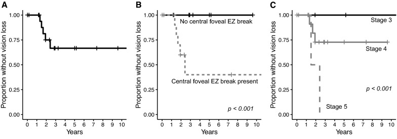
Results: A total of 20 eyes (11 patients) underwent cataract surgery and were followed for a median of 25.5 months (IQR 17.5-44.2 months). The median post-operative BCVA improvement was 10.5 letters (IQR 3.50-20.25). Nuclear sclerosis severity [β = 8.99 (95% CI 3.35, 14.6), p = 0.00177] was associated with post-operative change in BCVA and central foveal ellipsoid zone (EZ) breaks [OR 1.33 × 10⁻⁹ (95% CI 5.12 × 10⁻¹⁰-3.43 × 10⁻⁹), p < 0.001] on OCT was inversely correlated with post-operative BCVA > 20/40 using a multivariate generalized linear model. Central foveal EZ breaks [HR 1.77 × 10⁹ (95% CI 3.86 × 10⁸, 8.11 × 10⁹), p < 0.001] and MacTel Type 2 disease stage [HR 2.83, (95% CI 1.12, 7.12), p = 0.027] were independently associated with shorter time to vision loss of two lines or more in a multivariate Cox regression model.

Conclusions: Visual acuity significant improved after cataract surgery in eyes with MacTel Type 2 regardless of disease severity. The presence of central foveal EZ breaks may predict poorer post-operative visual acuity and subsequent vision loss from disease progression.

Keywords: Cataract surgery; Ellipsoid zone; Macular Telangiectasia.


Conflict of interest statement

The authors declare that they have no competing interests.

Figures

Fig. 1
Fig. 1
A Scatterplot showing best corrected visual acuity (BCVA) before and after cataract surgery. Pre-operative Snellen BCVA is plotted on the x-axis and post-operative Snellen BCVA is plotted on the y-axis. The diagonal line intersecting the origin represents no change in BCVA. Values above the diagonal line represent an increase in visual acuity and vice versa. B Dot and box plots showing post-op gain in ETDRS letters according to (left) nuclear sclerosis grade, (center) absence or presence of central foveal ellipsoid zone (EZ) break, and (right) MacTel disease stage
Fig. 2
Fig. 2
Kaplan–Meier curves of vision loss of 2 lines or more (10 or more ETDRS letters) in eyes with MacTel Type 2 after cataract surgery. A Kaplan–Meier curve of all eyes. B Kaplan–Meier curves of eyes with no central foveal ellipsoid zone (EZ) break (solid black line) and eyes with central foveal EZ break (dashed grey line). C Kaplan–Meier curves of eyes with MacTel Stage 3 (solid black line), Stage 4 (solid grey line) and Stage 5 (dashed grey line). p-values for univariate Cox regression are shown

References

    1. Powner MB, Gillies MC, Tretiach M, Scott A, Guymer RH, Hageman GS, et al. Perifoveal müller cell depletion in a case of macular telangiectasia type 2. Ophthalmology. 2010;117(12):2407–2416. doi: 10.1016/j.ophtha.2010.04.001. - DOI - PMC - PubMed
    1. Shen W, Fruttiger M, Zhu L, Chung SH, Barnett NL, Kirk JK, et al. Conditional Müller cell ablation causes independent neuronal and vascular pathologies in a novel transgenic model. J Neurosci. 2012;32(45):15715–15727. doi: 10.1523/JNEUROSCI.2841-12.2012. - DOI - PMC - PubMed
    1. Powner MB, Gillies MC, Zhu M, Vevis K, Hunyor AP, Fruttiger M. Loss of Müller’s cells and photoreceptors in macular telangiectasia type 2. Ophthalmology. 2013;120(11):2344–2352. doi: 10.1016/j.ophtha.2013.04.013. - DOI - PubMed
    1. Charbel Issa P, Gillies MC, Chew EY, Bird AC, Heeren TFC, Peto T, et al. Macular telangiectasia type 2. Prog Retin Eye Res. 2013;1(34):49–77. doi: 10.1016/j.preteyeres.2012.11.002. - DOI - PMC - PubMed
    1. Sallo FB, Peto T, Egan C, Wolf-Schnurrbusch UEK, Clemons TE, Gillies MC, et al. The IS/OS junction layer in the natural history of type 2 idiopathic macular telangiectasia. Invest Ophthalmol Vis Sci. 2012;53(12):7889–7895. doi: 10.1167/iovs.12-10765. - DOI - PMC - PubMed

LinkOut - more resources