. 2022 Sep;6(9):2441-2454.
doi: 10.1002/hep4.2018. Epub 2022 Jun 12.

A single hepatitis B virus genome with a reporter allows the entire viral life cycle to be monitored in primary human hepatocytes

Affiliations

A single hepatitis B virus genome with a reporter allows the entire viral life cycle to be monitored in primary human hepatocytes

Ariunaa Sumiyadorj et al. Hepatol Commun. 2022 Sep.

Abstract

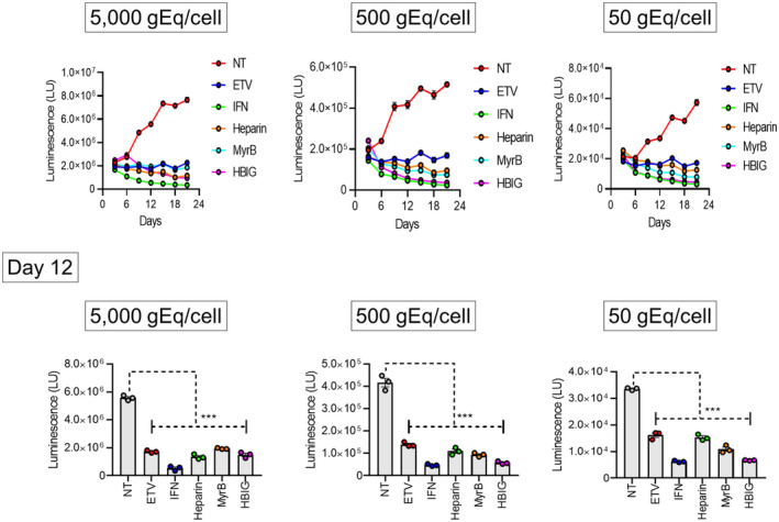
For the development of antiviral agents to eliminate hepatitis B virus (HBV), it is essential to establish an HBV cell culture system that can easily monitor HBV infection. Here, we created a novel HBV infection monitoring system using a luminescent 11-amino acid reporter, the high-affinity subunit of nano-luciferase binary technology (HiBiT). The HiBiT-coding sequence was inserted at the N-terminus of preS1 in a 1.2-fold plasmid encoding a genotype C HBV genome. After transfection of HepG2 cells with this HiBiT-containing plasmid, the supernatant was used to prepare a recombinant cell culture-derived virus (HiBiT-HBVcc). Primary human hepatocytes (PXB) were inoculated with HiBiT-HBVcc. Following inoculation, intracellular and extracellular HiBiT activity and the levels of various HBV markers were determined. Reinfection of naive PXB cells with HiBiT-HBVcc prepared from HiBiT-HBVcc-infected PXB cells was analyzed. When PXB cells were infected with HiBiT-HBVcc at several titers, extracellular HiBiT activity was detected in a viral titer-dependent manner and was correlated with intracellular HiBiT activity. Inhibitors of HBV entry or replication suppressed extracellular HiBiT activity. Viral DNA, RNA, and proteins were detectable, including covalently closed circular DNA, by Southern blot analysis. The synthesis of relaxed-circular DNA from single-stranded DNA in HiBiT-HBV decreased to one third of that of wild-type HBV, and the infectivity of HiBiT-HBVcc decreased to one tenth of that of wild-type HBVcc. HiBiT-HBVcc prepared from PXB cells harboring HiBiT-HBV was able to infect naive PXB cells. Conclusions: Recombinant HiBiT-HBV can undergo the entire viral life cycle, thus facilitating high-throughput screening for HBV infection in vitro using supernatants. This system will be a powerful tool for developing antiviral agents.


Conflict of interest statement

Nothing to report.

Figures

FIGURE 1
FIGURE 1
Insertion of the HiBiT‐coding sequence and detection of HiBiT activity following HiBiT‐HBVcc infection. (A) Two preS1 forms, a longer one and a shorter one, both of which have a myristoylation site at the N‐terminus, are produced from a wild‐type HBV genome. The HiBiT‐coding sequence was introduced into the N‐terminus of preS1 of a 1.2‐fold HBV genome. Thus, the region between amino acids 2 and 10 of the preS1 protein was replaced with HiBiT‐coding amino acids. In HiBiT‐HBV, the longer form does not have a myristoylation site owing to the insertion of the HiBiT‐coding sequence while the shorter one does have a myristoylation site. Numbering starts from the 5′‐end of the preS1 protein (upper, nucleotide) and the N‐terminus of the preS1 protein (lower, amino acid). (B) HepG2‐NTCP‐C4 cells were transfected with a plasmid containing the HiBiT‐coding sequence (pUC1.2×HBV‐C/HiBiT), and the medium was harvested and concentrated. Following quantification of HBV‐DNA levels, PXB cells were inoculated with medium containing HiBiT‐HBVcc at 50, 500, and 5000 gEq/cell. The medium was collected and replaced with fresh medium every 3 days until day 21, and extracellular HiBiT activity was measured (left panel). PXB cells were infected with HiBiT‐HBVcc at 500 gEq/cell, and the medium was collected and replaced with fresh medium every 3 days. Extracellular and intracellular HiBiT activity was measured every 3 days until day 18 (right panel). The data show the average HiBiT activity from three wells of a 96‐well plate (standard errors cannot be seen owing to their small values). Background HiBiT activity is shown as a dotted line. gEq/cell, genome equivalents per cell; HBV, hepatitis B virus; HBVcc, cell culture‐derived hepatitis B virus; HiBiT, high‐affinity nano‐luciferase binary technology; LU, light unit.
FIGURE 2
FIGURE 2
Specific increase of extracellular HiBiT activity after HiBiT‐HBVcc entry. PXB cells were infected with HiBiT‐HBVcc at 500 genome equivalents/cell. MyrB or heparin was added at the same time as infection or at day 3 after infection at the following concentrations: 2, 20, or 200 nm MyrB and 2, 20, or 200 IU/ml heparin. The medium was collected and replaced with fresh medium containing each compound every 3 days until day 12. (A) Schematic representation of the experiment. (B) Extracellular HiBiT activity was determined at day 12. Data show the average HiBiT activity from three wells of a 96‐well plate (with standard errors). Differences in the means between nontreatment and each treatment were analyzed with two‐way analysis of variance. HBV, hepatitis B virus; HBVcc, cell culture‐derived hepatitis B virus; heparin, heparin sodium; HiBiT, high‐affinity nano‐luciferase binary technology; LU, light unit; MyrB, myrcludex B. *p < 0.05, **p < 0.01, ***p < 0.001.
FIGURE 3
FIGURE 3
Comparison of various HBV markers between WT‐HBV and HiBiT‐HBV. (A) HepG2‐NTCP‐C4 cells were transfected with pUC1.2×HBV‐C/HiBiT or pUC1.2×HBV‐C, which lacks HiBiT insertion and was designated as WT. Medium was collected and precipitated by PEG, and PXB cells were infected with WT‐HBVcc or HiBiT‐HBVcc at 500 gEq/cell. Following WT‐HBVcc or HiBiT‐HBVcc infection, intracellular pgRNA and cccDNA levels; and extracellular HBV‐DNA, HBsAg (including large, middle, and small S protein), and HBcrAg levels were quantitated every 3 days until day 21 after infection. cccDNA levels are shown as copy number/100 ng total DNA. Additionally, pgRNA and β‐actin levels were determined by quantitative reverse transcription polymerase chain reaction, and pgRNA was normalized to β‐actin. Subsequently, the relative pgRNA levels were normalized to that of WT‐HBVcc at day 3, which was set to 1. Data show the average from three wells of a 96‐well plate (standard errors cannot be seen owing to their small values). (B) PXB cells were infected with WT‐HBVcc or HiBiT‐HBVcc at 50,000 gEq/ml, and cccDNA was extracted by Hirt's method. Southern blot analysis using a full‐length HBV probe was conducted on cccDNA extracted from the same number of cells. For assessing size markers for rcDNA, linearized DNA, and cccDNA, pUC19‐HBV, which is 3.2 kb in length, including a 540‐bp HBV sequence, was digested with Nb.BbvCI, ScaI, or no restriction enzyme and then subjected to Southern blot analysis. The full gel image with a shorter exposure time is shown in Figure S2. cccDNA, covalently closed circular DNA; gEq, genome equivalents; HBcrAg, hepatitis B virus core‐related antigen; HBsAg, hepatits B virus surface antigen; HBV, hepatitis B virus; HBV‐C, hepatitis B virus genotype C; HBVcc, cell culture‐derived hepatitis B virus; HiBiT, high‐affinity nano‐luciferase binary technology; NTCP, sodium taurocholate cotransporting polypeptide; PEG, polyethylene glycol; pgRNA, pregenomic RNA; PXB, primary human hepatocyte; rcDNA, relaxed circular DNA; WT, wild type.
FIGURE 4
FIGURE 4
Comparison of infectivity between WT‐HBV and HiBiT‐HBV. (A) The myristoylation site of the longer preS1 form, the second glycine, was substituted with alanine, and this mutant was designated G1A‐HBV. Thus, only the shorter preS1 form has a myristoylation site. The numbering starts from the 5′‐end of the preS1 protein (upper, nucleotide) and the N‐terminus of the preS1 protein (lower, amino acid). (B) PXB cells were infected with the indicated types of cell culture‐derived HBV, including WT‐, HiBiT‐, G1A‐, or G2A‐HBV, at 500, 5000, or 50,000 gEq/cell. At 15 days after infection, the cells were fixed and stained with DAPI and HBc. Scale bars, 200 μm. The numbers of DAPI‐ and HBc‐positive cells were counted in five different fields of view for each infection, using ImageJ; then, the numbers of DAPI‐ and HBc‐positive cells per HBV genome were calculated. DAPI, 4´,6‐diamidino‐2‐phenylindole; G1A, second glycine substituted with alanine; gEq, genome equivalents; HBc, hepatitis B virus core; HBV, hepatitis B virus; HiBiT, high‐affinity nano‐luciferase binary technology; PXB, primary human hepatocyte; WT, wild type.
FIGURE 5
FIGURE 5
Effect of antiviral compounds on HBV‐HiBiT infection. PXB cells were infected with HiBiT‐HBVcc at 50, 500, or 5000 gEq/cell, and 1 day later, each compound was added at the following concentration: 500 nm ETV, 1000 IU/ml IFN‐α2b, 100 IU/ml heparin, 20 nm MyrB, or 1 IU/ml human HBIG. The medium was collected and replaced with fresh medium containing each compound every 3 days until day 12. Extracellular HiBiT activity was determined using the collected media. The data show the average HiBiT activity from three wells of a 96‐well plate (with standard errors). The upper panel shows the time course of HiBiT activity, and the lower panel shows the results at day 12 for each level of HiBiT‐HBVcc infection. Differences in the means between NT and each treatment were analyzed with two‐way analysis of variance. ETV, entecavir; gEq, genome equivalents; HBIG, hepatitis B virus immunoglobulin; HBV, hepatitis B virus; HBVcc, cell culture‐derived hepatitis B virus; heparin, heparin sodium; HiBiT, high‐affinity nano‐luciferase binary technology; IFN, interferon; LU, light unit; MyrB, myrcludex B; NT, nontreatment; PXB, primary human hepatocyte. ***p < 0.001.
FIGURE 6
FIGURE 6
Cell‐free reinfection of PXB cells with HiBiT‐HBVcc. The supernatants from PXB cells harboring HiBiT‐HBV were collected and precipitated, and their HBV‐DNA levels were determined. Naive PXB cells were infected with HiBiT‐HBVcc at 500 genome equivalents/cell in the presence or absence of 20 nM MyrB. The medium was collected and replaced with fresh medium. (A) Schematic representation of the experiment. (B) Extracellular HiBiT activity was determined using medium collected every 3 days until day 15 after infection. At day 15, intracellular HiBiT activity was also determined. Data show the average HiBiT activity from three wells of a 96‐well plate (with standard errors). Upper panels show the time course results, and lower panels show the results at day 15. Statistical significance between two samples was examined by an unpaired t test; *p < 0.05, **p < 0.01. HBV, hepatitis B virus; HBVcc, cell culture‐derived hepatitis B virus; HiBiT, high‐affinity nano‐luciferase binary technology; LU, light unit; MyrB, myrcludex B; NT, nontreatment; PXB, primary human hepatocyte.
FIGURE 7
FIGURE 7
Iodixanol density gradient analysis of WT‐HBVcc and HiBiT‐HBVcc. (A) WT‐HBVcc or HiBiT‐HBVcc was concentrated and subjected to iodixanol density gradient analysis. Following ultracentrifugation, 14 fractions were collected from the top of the gradient and the density and HBV preS1 antigen (reflecting the large S protein), HBcrAg, and HBV‐DNA levels were measured for each fraction. PXB cells were infected with each fraction, and 12 days later, intracellular HBV‐DNA levels were determined and shown as copy number/100 ng total DNA. Data show the average from three wells of a 96‐well plate (with standard errors). (B) Fractions containing the highest HBV‐DNA levels from WT‐HBV and HiBiT‐HBV were used to take images of virus particles by negative‐staining electron microscopy. Left panel shows a Dane particle of WT‐HBV; right panel shows one of HiBiT‐HBV. Scale bars, 50 nm. HBcrAg, hepatitis B virus core‐related antigen; HBV, hepatitis B virus; HBVcc, cell culture‐derived hepatitis B virus; HiBiT, high‐affinity nano‐luciferase binary technology; NT, nontreatment; PXB, primary human hepatocyte; WT, wild type.

Similar articles

Cited by

References

    1. Polaris Observatory Collaborators . Global prevalence, treatment, and prevention of hepatitis B virus infection in 2016: a modelling study. Lancet Gastroenterol Hepatol. 2018;3:383–403. - PubMed
    1. Muller B, Daecke J, Fackler OT, Dittmar MT, Zentgraf H, Krausslich HG. Construction and characterization of a fluorescently labeled infectious human immunodeficiency virus type 1 derivative. J Virol. 2004;78:10803–13. - PMC - PubMed
    1. Terahara K, Yamamoto T, Mitsuki YY, Shibusawa K, Ishige M, Mizukoshi F, et al. Fluorescent reporter signals, EGFP, and DsRed, encoded in HIV‐1 facilitate the detection of productively infected cells and cell‐associated viral replication levels. Front Microbiol. 2012;2:280. - PMC - PubMed
    1. Moradpour D, Evans MJ, Gosert R, Yuan Z, Blum HE, Goff SP, et al. Insertion of green fluorescent protein into nonstructural protein 5A allows direct visualization of functional hepatitis C virus replication complexes. J Virol. 2004;78:7400–9. - PMC - PubMed
    1. Shimakami T, Welsch C, Yamane D, McGivern DR, Yi M, Zeuzem S, et al. Protease inhibitor‐resistant hepatitis C virus mutants with reduced fitness from impaired production of infectious virus. Gastroenterology. 2011;140:667–75. - PMC - PubMed

Publication types