

Rhino-orbito-cerebral mucormycosis caused by Rhizopus microsporus var. microsporus in a diabetic patient with COVID-19
- PMID: 35691738
- PMCID: PMC9181896
- DOI: 10.1016/j.abd.2022.02.001
Rhino-orbito-cerebral mucormycosis caused by Rhizopus microsporus var. microsporus in a diabetic patient with COVID-19
Abstract
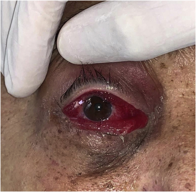
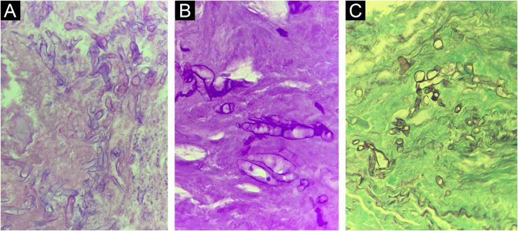
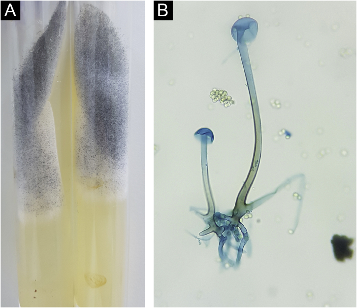
COVID-19 disease caused by the SARS-CoV-2 coronavirus causes a wide range of clinical manifestations, ranging from mild to severe, with the main ones affecting the respiratory tract, such as pneumonia. In patients with greater severity, the high frequency of bacterial and fungal coinfection stands out, a situation related both to the patient's pre-existing comorbidities and due to the hospitalization itself. Cases of mucormycosis associated with COVID-19 were highlighted in the lay and scientific media, with the increase in mycosis cases being directly and indirectly attributed to the viral infection. This report describes a case of rhino-orbito-cerebral mucormycosis in a diabetic patient hospitalized for COVID-19, whose diagnosis was confirmed by identifying the agent Rhizopus microsporus var. microsporus through culture for fungi and PCR examination.
Keywords: Amphotericin B; COVID-19; Coronavirus; Diabetes mellitus; Diagnosis; Mucormycosis.
Copyright © 2022 Sociedade Brasileira de Dermatologia. Published by Elsevier España, S.L.U. All rights reserved.
Figures
Similar articles
-
Rhizopus microsporus as Causative Agent of Mucormycosis in COVID-19 Patient.Clin Lab. 2022 Dec 1;68(12). doi: 10.7754/Clin.Lab.2022.220232. Clin Lab. 2022. PMID: 36546751
-
Mixed mold infection with Aspergillus fumigatus and Rhizopus microsporus in a severe acute respiratory syndrome Coronavirus 2 (SARS-CoV-2) patient.Infect Dis Now. 2021 Oct;51(7):633-635. doi: 10.1016/j.idnow.2021.01.010. Epub 2021 Jan 27. Infect Dis Now. 2021. PMID: 33527098 Free PMC article. No abstract available.
-
First report of COVID-19-associated rhino-orbito-cerebral mucormycosis in pediatric patients with type 1 diabetes mellitus.J Mycol Med. 2021 Dec;31(4):101203. doi: 10.1016/j.mycmed.2021.101203. Epub 2021 Sep 4. J Mycol Med. 2021. PMID: 34517273 Free PMC article.
-
COVID-19-associated rhino-orbital-cerebral mucormycosis: A systematic review, meta-analysis, and meta-regression analysis.Indian J Pharmacol. 2021 Nov-Dec;53(6):499-510. doi: 10.4103/ijp.ijp_839_21. Indian J Pharmacol. 2021. PMID: 34975140 Free PMC article.
-
Rhino-orbital-cerebral-mucormycosis in COVID-19: A systematic review.Indian J Pharmacol. 2021 Jul-Aug;53(4):317-327. doi: 10.4103/ijp.ijp_419_21. Indian J Pharmacol. 2021. PMID: 34414911 Free PMC article.
Cited by
-
Increase in mucormycosis hospitalizations in southeastern Brazil during the COVID-19 pandemic: a 2010-2021 time series.Rev Soc Bras Med Trop. 2023 Feb 20;56:e0333. doi: 10.1590/0037-8682-0333-2022. eCollection 2023. Rev Soc Bras Med Trop. 2023. PMID: 36820656 Free PMC article.
References
-
- Cornely O.A., Alastruey-Izquierdo A., Arenz D., Chen S.C.A., Dannaoui E., Hochhegger B., et al. Mucormycosis ECMM MSG Global Guideline Writing Group Global guideline for the diagnosis and management of mucormycosis: an initiative of the European Confederation of Medical Mycology in cooperation with the Mycoses Study Group Education and Research Consortium. Lancet Infect Dis. 2019;19:e405–e421. - PMC - PubMed
Publication types
MeSH terms
Substances
Supplementary concepts
LinkOut - more resources
Full Text Sources
Medical
Miscellaneous
