Review
. 2022 Sep;45(9):678-691.
doi: 10.1016/j.tins.2022.05.007. Epub 2022 Jun 9.

Neural consequences of chronic sleep disruption

Affiliations
Review

Neural consequences of chronic sleep disruption

Zachary Zamore et al. Trends Neurosci. 2022 Sep.

Abstract

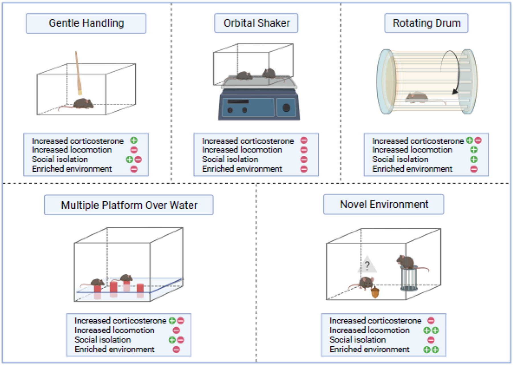
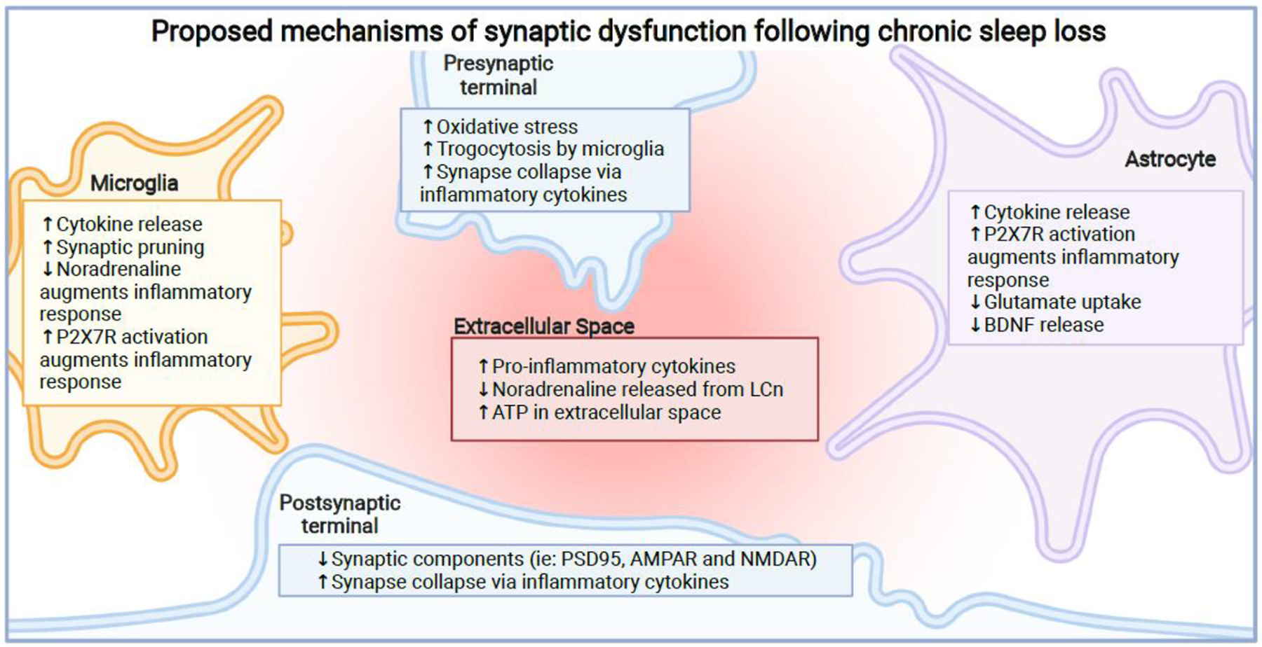
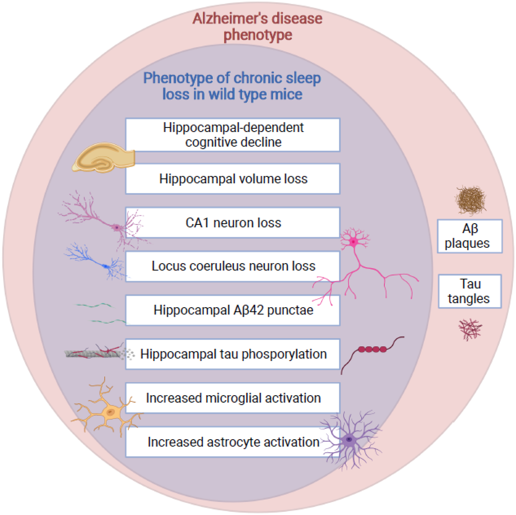
Recent studies in both humans and animal models call into question the completeness of recovery after chronic sleep disruption. Studies in humans have identified cognitive domains particularly vulnerable to delayed or incomplete recovery after chronic sleep disruption, including sustained vigilance and episodic memory. These findings, in turn, provide a focus for animal model studies to critically test the lasting impact of sleep loss on the brain. Here, we summarize the human response to sleep disruption and then discuss recent findings in animal models examining recovery responses in circuits pertinent to vigilance and memory. We then propose pathways of injury common to various forms of sleep disruption and consider the implications of this injury in aging and in neurodegenerative disorders.

Keywords: amyloid; locus coeruleus; neurodegeneration; sleep deprivation; sleep fragmentation; sleep restriction.


Conflict of interest statement

Declaration of interests The authors declare no competing interests.

Figures

Fig 1.
Fig 1.
Chronic sleep disruption (CSD) paradigms. Approaches to chronic disruption in animal models vary not only by the way in which sleep is disrupted, but also by environment (including stress), intermittency or constancy of wakefulness, elicited motor activity and learning opportunities. Shown are specific attributes and potential confounders that may influence results. For each CSD paradigm, phenomena that were observed in multiple studies are indicated by a green plus sign, and phenomena that were absent in some or most of the relevant studies are indicated by a red negative sign.
Fig 2.
Fig 2.
Duration-dependent effects of sleep loss on locus coeruleus neurons (LCn). Short-term wakefulness (awake for three consecutive hours across the habitual sleep (lights-on) period) upregulates mitochondrial sirtuin type 3 (SirT3) activity, which then results in nuclear translocation of FoxO3a and transcriptional activation of anti-oxidants and PGC-1α to enhance mitochondrial biogenesis. In contrast, extended wakefulness (for 8 hours a day in the lights-on period for three consecutive days) reduces SirT3 protein and its NAD+-synthesizing enzymes, thereby reducing SirT3 activity and increasing mitochondrial acetylation of (and thereby inactivating) anti-oxidant enzymes, electron transport chain proteins, and FoxO3a. Mechanisms by which LCn switch from an adaptive to maladaptive response are not known.
Fig 3.
Fig 3.
Proposed mechanisms of synaptic dysfunction following chronic sleep loss. Chronic sleep loss impairs the functionality of the synapse, with consequences seen in both the pre- and post-synapse in addition to the surrounding glial cells. With most of these effects seen in the hippocampus or cortex, these effects likely impair learning and memory. Microglia can release cytokines contributing to the inflammatory environment[105]. The loss of LCn leads to loss of anti-inflammatory effects of noradrenaline and increased microglial-mediated synaptic pruning[,–95]. Elevated levels of ATP in the extracellular space activates microglial P2X7 receptors, amplifying inflammation[81,82]. In astrocytes, there is an increase in cytokine production contributing to the inflammatory environment[83]. Since astrocytes also express P2X7 receptors, elevated ATP in the extracellular space activates these receptors and amplifies the inflammatory response[80,82]. In addition, the ability of astrocytes to regulate glutamate levels in the synapse is impaired, which can lead to neuronal excitotoxicity [90]. Astrocytes also release less BDNF, which may contribute to synapse loss[84]. In the extracellular space, there is an increase in cytokines[83], a decrease in noradrenaline likely due to LCn loss, and an increase in ATP[80]. In the pre-synapse, there is increased oxidative stress[80], increase in inappropriately-timed trogocytosis [–95] and inflammation-mediated synaptic loss[85,86]. Finally, in the post-synapse, there is loss of synaptic components due to synapse loss[80], which may be mediated by the inflammatory environment [85,86]. In summary, chronic sleep loss creates a pro-inflammatory environment at the synapse level which is characterized by impaired glial functionality and synapse loss.
Fig 4.
Fig 4.
Phenotypic overlap between chronic sleep disruption and Alzheimer’s disease. Commonalities in the neural response to chronic sleep disruption in wild type mice are shown as a subset of the neural and cognitive changes observed in Alzheimer’s disease. Of note, wild type mice do not naturally develop amyloid plaques or tau tangles. In animal models with expression of human amyloid precursor protein and/or tau mutations observed in familial degenerative disorders, sleep disruption increases plaque and tangle formation, as well as tau propagation.

Similar articles

Cited by

References

    1. Dinges DF et al. (1997) Cumulative sleepiness, mood disturbance, and psychomotor vigilance performance decrements during a week of sleep restricted to 4–5 hours per night. Sleep 20, 267–277 - PubMed
    1. Van Dongen HP et al. (2003) The cumulative cost of additional wakefulness: dose-response effects on neurobehavioral functions and sleep physiology from chronic sleep restriction and total sleep deprivation. Sleep 26, 117–126 - PubMed
    1. Belenky G et al. (2003) Patterns of performance degradation and restoration during sleep restriction and subsequent recovery: a sleep dose-response study. J Sleep Res 12, 1–12. 337 [pii] - PubMed
    1. Banks S et al. (2010) Neurobehavioral dynamics following chronic sleep restriction: dose-response effects of one night for recovery. Sleep 33, 1013–1026. 10.1093/sleep/33.8.1013 - DOI - PMC - PubMed
    1. Pejovic S et al. (2013) Effects of recovery sleep after one work week of mild sleep restriction on interleukin-6 and cortisol secretion and daytime sleepiness and performance. Am J Physiol Endocrinol Metab 305, E890–896. 10.1152/ajpendo.00301.2013 - DOI - PMC - PubMed

Publication types