Integrated Behavioral, Genetic and Brain Circuit Visualization Methods to Unravel Functional Anatomy of Zebrafish Amygdala
- PMID: 35692259
- PMCID: PMC9174433
- DOI: 10.3389/fnana.2022.837527
Integrated Behavioral, Genetic and Brain Circuit Visualization Methods to Unravel Functional Anatomy of Zebrafish Amygdala
Abstract
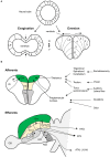
The mammalian amygdala is a complex forebrain structure consisting of a heterogeneous group of nuclei derived from the pallial and subpallial telencephalon. It plays a critical role in a broad range of behaviors such as emotion, cognition, and social behavior; within the amygdala each nucleus has a distinct role in these behavioral processes. Topological, hodological, molecular, and functional studies suggest the presence of an amygdala-like structure in the zebrafish brain. It has been suggested that the pallial amygdala homolog corresponds to the medial zone of the dorsal telencephalon (Dm) and the subpallial amygdala homolog corresponds to the nuclei in the ventral telencephalon located close to and topographically basal to Dm. However, these brain regions are broad and understanding the functional anatomy of the zebrafish amygdala requires investigating the role of specific populations of neurons in brain function and behavior. In zebrafish, the highly efficient Tol2 transposon-mediated transgenesis method together with the targeted gene expression by the Gal4-UAS system has been a powerful tool in labeling, visualizing, and manipulating the function of specific cell types in the brain. The transgenic resource combined with neuronal activity imaging, optogenetics, pharmacology, and quantitative behavioral analyses enables functional analyses of neuronal circuits. Here, we review earlier studies focused on teleost amygdala anatomy and function and discuss how the transgenic resource and tools can help unravel the functional anatomy of the zebrafish amygdala.
Keywords: Gal4-UAS system; amygdala; dorsomedial telencephalon; emotion; pallium; subpallium; transgenic; zebrafish.
Copyright © 2022 Lal and Kawakami.
Conflict of interest statement
The authors declare that the research was conducted in the absence of any commercial or financial relationships that could be construed as a potential conflict of interest.
Figures


Similar articles
-
Identification of a neuronal population in the telencephalon essential for fear conditioning in zebrafish.BMC Biol. 2018 Apr 25;16(1):45. doi: 10.1186/s12915-018-0502-y. BMC Biol. 2018. PMID: 29690872 Free PMC article.
-
The Everted Amygdala of Ray-Finned Fish: Zebrafish Makes a Case.Brain Behav Evol. 2022;97(6):321-335. doi: 10.1159/000525669. Epub 2022 Jun 27. Brain Behav Evol. 2022. PMID: 35760049 Review.
-
The organization of the zebrafish pallium from a hodological perspective.J Comp Neurol. 2022 Jun;530(8):1164-1194. doi: 10.1002/cne.25268. Epub 2021 Nov 11. J Comp Neurol. 2022. PMID: 34697803
-
Evolution and Development of Amygdala Subdivisions: Pallial, Subpallial, and Beyond.Brain Behav Evol. 2023;98(1):1-21. doi: 10.1159/000527512. Epub 2022 Oct 20. Brain Behav Evol. 2023. PMID: 36265454 Review.
-
Dorsomedial telencephalon of lungfishes: a pallial or subpallial structure? Criteria based on histology, connectivity, and histochemistry.J Comp Neurol. 1990 Apr 1;294(1):14-29. doi: 10.1002/cne.902940103. J Comp Neurol. 1990. PMID: 2324329
Cited by
-
Genetic Identification of Neural Circuits Essential for Active Avoidance Fear Conditioning in Adult Zebrafish.Methods Mol Biol. 2024;2707:169-181. doi: 10.1007/978-1-0716-3401-1_11. Methods Mol Biol. 2024. PMID: 37668912
-
Distinct acute stressors produce different intensity of anxiety-like behavior and differential glutamate release in zebrafish brain.Front Behav Neurosci. 2024 Oct 23;18:1464992. doi: 10.3389/fnbeh.2024.1464992. eCollection 2024. Front Behav Neurosci. 2024. PMID: 39508031 Free PMC article.
-
Embryonic Zebrafish as a Model for Investigating the Interaction between Environmental Pollutants and Neurodegenerative Disorders.Biomedicines. 2024 Jul 13;12(7):1559. doi: 10.3390/biomedicines12071559. Biomedicines. 2024. PMID: 39062132 Free PMC article. Review.
-
Molecular organization of neuronal cell types and neuromodulatory systems in the zebrafish telencephalon.Curr Biol. 2024 Jan 22;34(2):298-312.e4. doi: 10.1016/j.cub.2023.12.003. Epub 2023 Dec 28. Curr Biol. 2024. PMID: 38157860 Free PMC article.
-
Does a Vertebrate Morphotype of Pallial Subdivisions Really Exist?Brain Behav Evol. 2024;99(4):230-247. doi: 10.1159/000537746. Epub 2024 Jul 16. Brain Behav Evol. 2024. PMID: 38952102 Free PMC article. Review.
References
Publication types
LinkOut - more resources
Full Text Sources
Research Materials

