. 2022 May 26:9:853819.
doi: 10.3389/fvets.2022.853819. eCollection 2022.

Tissue Expression Analysis, Cloning, and Characterization of the 5'-Regulatory Region of the Bovine LATS 1 Gene

Affiliations

Tissue Expression Analysis, Cloning, and Characterization of the 5'-Regulatory Region of the Bovine LATS 1 Gene

Dawei Wei et al. Front Vet Sci. .

Abstract

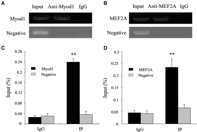
As a member of the large tumor suppressor (LATS) gene family, LATS1 plays an important role in regulating muscle growth and development. In this study, we determined the distinct exhibit patterns of tissue expression of bovine LATS1. Further, we determined the functional proximal minimal promoter of bovine LATS1 and identified the key transcription factors in the core promoter region to elucidate its molecular regulation mechanism. The results showed that bovine LATS1 was highly expressed in the longissimus thoracis and upregulation in infancy muscle. An electrophoretic mobility shift assay (EMSA) and chromatin immunoprecipitation (ChIP) assay in combination with site-directed mutation and small interfering RNA (siRNA) interference demonstrated that myogenic differentiation 1 (Myod1) and myocyte enhancer factor 2A (MEF2A) binding in the core promoter region (-298/-123 bp) play important roles in the transcriptional regulation of the bovine LATS1 promoter. Taken together, these interactions provide insight into the regulatory mechanisms of LATS1 transcription in mediating skeletal muscle growth in cattle.

Keywords: LATS1 gene; core promoter; expression; factor; transcription.


Conflict of interest statement

The authors declare that the research was conducted in the absence of any commercial or financial relationships that could be construed as a potential conflict of interest. The reviewer CM declared a shared affiliation with the author SR to the handling editor at the time of the review.

Figures

Figure 1
Figure 1
Expression pattern analysis of the bovine large tumor suppressor 1 (LATS1). (A) Analysis of the bovine LATS1 expression pattern in tissues and organs. (B) Expression pattern of the bovine LATS1 messenger RNA (mRNA) in longissimus thoracis at different developmental stages. The value of each column represents the mean ± SD of three independent experiments. Different uppercases and lowercases represent p < 0.01 and p < 0.05, respectively.
Figure 2
Figure 2
Structural characteristics of the bovine LATS1 gene. (A) The detailed genomic, mRNA, and protein components are containing the 5′/3′-untranslated region (5′/3′-UTR) and the open reading frame (ORF). (B) 5′-regulatory region sequence of the bovine LATS1 gene. Arrows mark the transcription initiation sites. The transcription factor binding sites are boxed. (C) Dashed lines indicate the GC percentage as represented on the y-axis and the x-axis denotes the bp position in the 5'-untranslated region. Coordinates are given relative to the translational start site (shown as + 1).
Figure 3
Figure 3
The construction of LATS1 protein phylogenetic tree. On the left side, there is the clustering of LATS1 protein in different species and on the right side, there is the architecture and characteristics of LATS1 protein amino acid length in corresponding species.
Figure 4
Figure 4
Isolation and analysis of the functional proximal minimal promoter of LATS1. A series of plasmids containing 5' unidirectional deletions of the promoter region of the LATS1 gene fused in-frame to the luciferase gene were transfected into myoblasts and myotube cells. The results are expressed as the means ± SD in arbitrary units based on the firefly/Renilla luciferase activity in triplicate transfections. “*” p < 0.05 and “**” p < 0.01.
Figure 5
Figure 5
Analysis of transcriptional activity for the corresponding transcription factor in Qinchuan cattle myoblast cells (QCMCs). (A) Site-directed mutagenesis for homeobox A5 (HOXA5), myogenic differentiation 1 (Myod1), forkhead box O1 (FoxO1), and myocyte enhancer factor 2A (MEF2A) sites were carried out in the construct pGL−298/+167. The correspondence constructs were transiently transfected into QCMCs and pGL−123/+167 construct was used as a negative control. (B) Myod1 and MEF2A knockdown by small interfering RNA (siRNA) co-transfected with pGL−298/+167 in QCMCs. The NC siRNA was used as a negative control. The results are expressed as the means ± SD in arbitrary units based on the firefly/Renilla luciferase activity for triplicate transfections. (C) Multialignment sequence analysis of the Myod1 and MEF2A transcription factor binding sites in the promoter region of the LATS1 gene in cattle, sheep, horse, and goats. “*” p < 0.05 and “**” p < 0.01.
Figure 6
Figure 6
Identification of Myod1 and MEF2A bind to the proximal minimal promoter of LATS1 by electrophoretic mobility shift assay (EMSA) in vitro. (A,B) Nuclear protein extracts were incubated with 5′-biotin-labeled probe containing the Myod1- and MEF2A-binding site in the presence or absence of competitor (lane 2), 50X mutation probe (lane 3), and 50X unlabeled probes (lane 4). The supershift assay was conducted using 10 μg of anti-Myod1 and anti-MEF2A antibodies (lane 5). The arrows mark the main complexes and supershifted, respectively.
Figure 7
Figure 7
Chromatin immunoprecipitation (ChIP) assay of Myod1 and MEF2A binding to the LATS1 promoter in vivo. ChIP-PCR products of Myod1 (A) and MEF2A (B) input and immunoprecipitated were amplified and identified by 1% agarose electrophoresis from muscle. ChIP-qPCR assays detected the enrichment of DNA fragments in samples immunoprecipitated with Myod1 (C) and MEF2A (D) antibodies. We used the input was total chromatin from muscle, while the products from rabbit antiimmunoglobulin G (IgG) and an intragenic DNA fragment of LATS1 exon 3. “**” p < 0.01.
Figure 8
Figure 8
A proposed schematic summary of the regulation of LATS1 expression by Myod1 and MEF2A transcription factors (TFs).

References

    1. Li XZ, Yan CG, Zan LS. Current situation and future prospects for beef production in China—a review. Asian-Australas J Anim Sci. (2018) 31:984–91. 10.5713/ajas.18.0212 - DOI - PMC - PubMed
    1. Lecker SH, Jagoe RT, Gilbert A, Gomes M, Baracos V, Bailey J, et al. . Multiple types of skeletal muscle atrophy involve a common program of changes in gene expression. FASEB J. (2004) 18:39–51. 10.1096/fj.03-0610com - DOI - PubMed
    1. Fiszman MY. [Molecular strategies involved in the control of gene expression during differentiation of muscle cells]. Reprod Nutr Dev. (1988) 28:703–13. 10.1051/rnd:19880504 - DOI - PubMed
    1. Listrat A, Lebret B, Louveau I, Astruc T, Bonnet M, Lefaucheur L, et al. . How muscle structure and composition influence meat and flesh quality. Sci World J. (2016) 2016:3182746. 10.1155/2016/3182746 - DOI - PMC - PubMed
    1. Yi J, Lu L, Yanger K, Wang W, Sohn BH, Stanger BZ, et al. . Large tumor suppressor homologs 1 and 2 regulate mouse liver progenitor cell proliferation and maturation through antagonism of the coactivators YAP and TAZ. Hepatology. (2016) 64:1757–72. 10.1002/hep.28768 - DOI - PMC - PubMed