

Prevalence and Clinicopathologic Features of Canine Metastatic Melanoma Involving the Central Nervous System: A Retrospective Analysis and Comparative Review
- PMID: 35692802
- PMCID: PMC9186031
- DOI: 10.3389/fonc.2022.868004
Prevalence and Clinicopathologic Features of Canine Metastatic Melanoma Involving the Central Nervous System: A Retrospective Analysis and Comparative Review
Abstract
Background: Central nervous system (CNS) involvement is the leading cause of death in malignant melanoma. Rodent models, while vital to mechanistic investigation, have had limited success identifying effective therapies for melanoma brain metastases. The companion dog with de novo melanoma is a promising complementary model for developmental therapeutic investigation, as these tumors occur in an immunologically outbred host that has shared environmental exposures with humans. However, relatively little is known regarding the prevalence and clinicopathological features of canine melanoma metastasis to the CNS. To further validate the dog as an appropriate model for human metastatic melanoma, the aims of this study were to determine the rate of CNS metastasis and associated clinicopathologic features in canine malignant melanoma.
Methods: Medical records of dogs diagnosed with malignant melanoma from 1985-2019 at the University of California Davis Veterinary Medical Teaching Hospital were assessed retrospectively. Clinicopathologic features were compared between dogs with CNS metastasis (CNS+) and dogs without CNS metastasis (CNS-). Site of CNS involvement and associated neurological signs were analyzed via Wilcoxon-Mann-Whitney rank sum and Fisher's exact tests. Survival data were analyzed via Kaplan-Meier estimates.
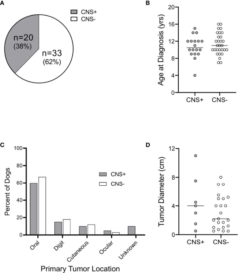
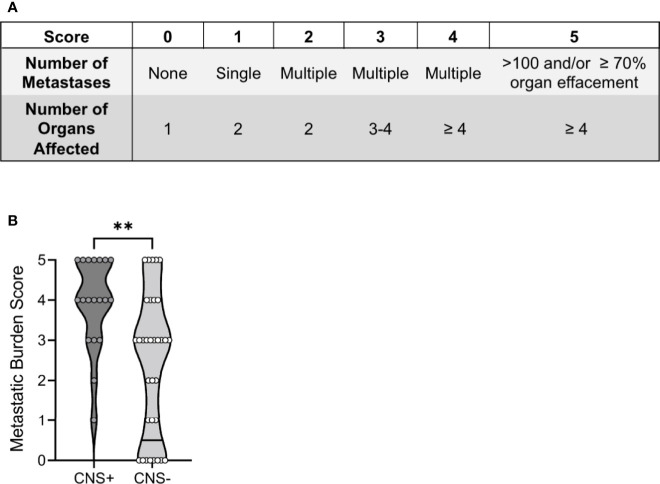
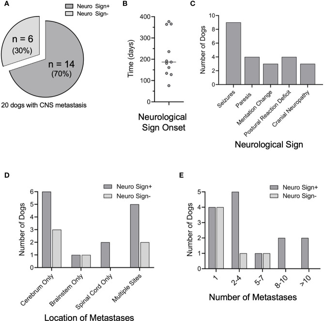
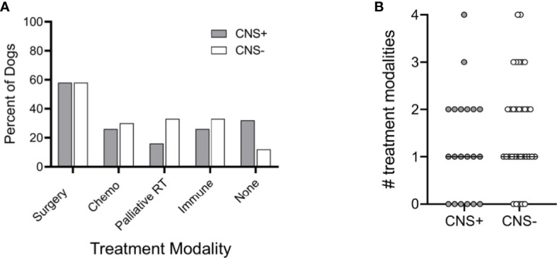
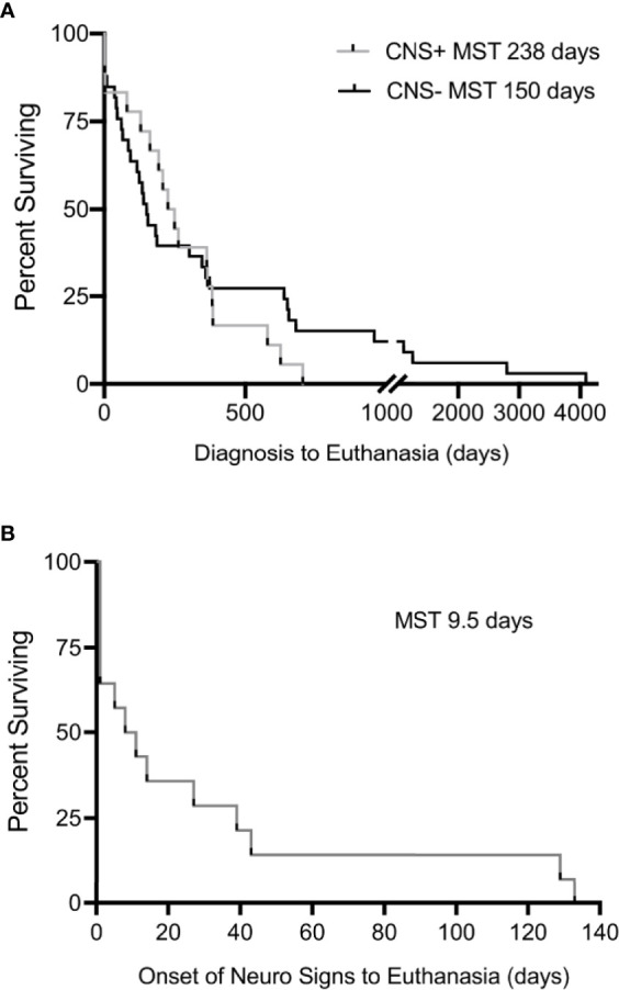
Results: CNS metastasis was identified in 38% of dogs in this study (20/53). The oral cavity was the most common site of primary melanoma in both groups [CNS+: n=12 (60%) vs. CNS-: n=22 (67%); p>0.99]. The total burden of metastatic disease was higher in the CNS+ group (CNS+: 4, 95% CI 3-5 vs. CNS-: 3, 95% CI 1-3; p<0.001). The cerebrum was the most common site of CNS metastasis (n=15, 75%) and seizures were the most observed neurological sign (n=9, 64%). There was no difference in overall survival between CNS+ and CNS- groups. However, the median survival time following onset of neurological signs was 9.5 days (95% CI 1-43), with 5 dogs euthanized within 24 hours of the onset of neurological signs.
Conclusions: Canine and human MM patients share similar rates of CNS metastasis and clinical presentation. This study will guide clinical management of canines with malignant melanoma and inform future studies using dogs with spontaneously occurring melanoma as a preclinical model for human melanoma brain metastases.
Keywords: brain; canine; central nervous system; large-animal model; malignant melanoma; seizure.
Copyright © 2022 Razmara, Wittenburg, Al-Nadaf, Toedebusch, Meyers and Toedebusch.
Conflict of interest statement
The authors declare that the research was conducted in the absence of any commercial or financial relationships that could be construed as a potential conflict of interest.
Figures
Similar articles
-
A retrospective analysis of 140 dogs with oral melanoma treated with external beam radiation.Vet Radiol Ultrasound. 2003 May-Jun;44(3):352-9. doi: 10.1111/j.1740-8261.2003.tb00468.x. Vet Radiol Ultrasound. 2003. PMID: 12816381
-
Treatment patterns and clinical outcomes for patients with melanoma and central nervous system metastases: A real-world study.Cancer Med. 2022 Jan;11(1):139-150. doi: 10.1002/cam4.4438. Epub 2021 Dec 7. Cancer Med. 2022. PMID: 34874127 Free PMC article.
-
Predictive factors for the development of brain metastasis in advanced unresectable metastatic melanoma.Am J Clin Oncol. 2011 Dec;34(6):603-10. doi: 10.1097/COC.0b013e3181f9456a. Am J Clin Oncol. 2011. PMID: 21150567
-
The treatment of brain metastases from malignant melanoma.Semin Oncol. 2002 Oct;29(5):518-24. doi: 10.1053/sonc.2002.35247. Semin Oncol. 2002. PMID: 12407517 Review.
-
Review on Canine Oral Melanoma: An Undervalued Authentic Genetic Model of Human Oral Melanoma?Vet Pathol. 2021 Sep;58(5):881-889. doi: 10.1177/0300985821996658. Epub 2021 Mar 9. Vet Pathol. 2021. PMID: 33685309 Review.
Cited by
-
Metastatic extradural melanoma of the lumbar spine in a cat.Vet Med Sci. 2023 Nov;9(6):2393-2398. doi: 10.1002/vms3.1248. Epub 2023 Sep 1. Vet Med Sci. 2023. PMID: 37656442 Free PMC article.
-
Diagnostic immunohistochemistry of primary and secondary central nervous system neoplasms of dogs and cats.J Vet Diagn Invest. 2024 Mar;36(2):153-168. doi: 10.1177/10406387231221858. Epub 2024 Jan 17. J Vet Diagn Invest. 2024. PMID: 38234003 Free PMC article. Review.
-
Case report: MRI and CT imaging features of a melanocytic tumour affecting a cervical vertebra in an adult dog, and review of differential diagnosis for T1W-hyperintense lesions.Front Vet Sci. 2024 Apr 9;11:1334813. doi: 10.3389/fvets.2024.1334813. eCollection 2024. Front Vet Sci. 2024. PMID: 38655532 Free PMC article.