. 2022 May 27:12:887546.
doi: 10.3389/fonc.2022.887546. eCollection 2022.

Multimodal MRI-Based Radiomics-Clinical Model for Preoperatively Differentiating Concurrent Endometrial Carcinoma From Atypical Endometrial Hyperplasia

Affiliations

Multimodal MRI-Based Radiomics-Clinical Model for Preoperatively Differentiating Concurrent Endometrial Carcinoma From Atypical Endometrial Hyperplasia

Jieying Zhang et al. Front Oncol. .

Abstract

Objectives: To develop and validate a radiomics model based on multimodal MRI combining clinical information for preoperative distinguishing concurrent endometrial carcinoma (CEC) from atypical endometrial hyperplasia (AEH).

Materials and methods: A total of 122 patients (78 AEH and 44 CEC) who underwent preoperative MRI were enrolled in this retrospective study. Radiomics features were extracted based on T2-weighted imaging (T2WI), diffusion-weighted imaging (DWI), and apparent diffusion coefficient (ADC) maps. After feature reduction by minimum redundancy maximum relevance and least absolute shrinkage and selection operator algorithm, single-modal and multimodal radiomics signatures, clinical model, and radiomics-clinical model were constructed using logistic regression. Receiver operating characteristic (ROC) analysis, calibration curves, and decision curve analysis were used to assess the models.

Results: The combined radiomics signature of T2WI, DWI, and ADC maps showed better discrimination ability than either alone. The radiomics-clinical model consisting of multimodal radiomics features, endometrial thickness >11mm, and nulliparity status achieved the highest area under the ROC curve (AUC) of 0.932 (95% confidential interval [CI]: 0.880-0.984), bootstrap corrected AUC of 0.922 in the training set, and AUC of 0.942 (95% CI: 0.852-1.000) in the validation set. Subgroup analysis further revealed that this model performed well for patients with preoperative endometrial biopsy consistent and inconsistent with postoperative pathologic data (consistent group, F1-score = 0.865; inconsistent group, F1-score = 0.900).

Conclusions: The radiomics model, which incorporates multimodal MRI and clinical information, might be used to preoperatively differentiate CEC from AEH, especially for patients with under- or over-estimated preoperative endometrial biopsy.

Keywords: endometrial hyperplasia; endometrial neoplasms; magnetic resonance imaging; radiomics; texture analysis.


Conflict of interest statement

Author LX was employed by GE Healthcare. The remaining authors declare that the research was conducted in the absence of any commercial or financial relationships that could be construed as a potential conflict of interest.

Figures
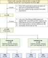
Figure 1
Figure 1
Flowchart of patient enrollment in this study.
Figure 2
Figure 2
Workflow of radiomic analysis. (A) MR imaging segmentation. Three-dimensional (3D) segmentation of tumors in MR images. (B) Radiomic feature extraction. Radiomic features, including shape, intensity, and texture, were extracted from the tumor volume. (C) Feature selection process. The stability analysis, the minimum redundancy maximum relevance (mRMR), and the least absolute shrinkage and selection operator (LASSO) algorithm were used for the radiomic feature selection. (D) Model construction. Radiomics signatures were constructed using a binary logistic regression model. Finally, a nomogram for the optimal model was developed. (E) Model assessment. The performances of our models were evaluated by discrimination, calibration, and clinical utility, as well as subgroup analysis. VOI, volume of interest; GLCM, gray level co-occurrence matrix; GLSZM, gray level size zone matrix; GLRLM, gray level run length matrix; GLDM, gray level dependence matrix.
Figure 3
Figure 3
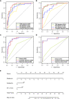
ROCs of the four radiomics signatures in the training (A) and validation (B) sets. ROCs of the clinical model, radiomics signature, and radiomics-clinical model in the training (C) and validation sets (D). (E) Preoperative nomogram of the radiomics-clinical model. ET, endometrial thickness.
Figure 4
Figure 4
The calibration plots of the radiomics-clinical model in the training (A) and validation sets (B). Patient risk scores output by the radiomics-clinical model in the training (C) and validation sets (D), while orange bars show scores for those who have concurrent endometrial carcinoma.
Figure 5
Figure 5
Decision curve analysis for the models in the validation set. It can be concluded that when the threshold probability is over 30% approximately, the radiomics-clinical model could provide extra profits over the “treat-all” or “treat-none” scheme, the combined radiomics signature, and the clinical model.
Figure 6
Figure 6
(A) Heatmap showing the models’ performance in the subgroups of patients with preoperative endometrial biopsy consistent and inconsistent with postoperative pathologic data. A deeper red indicates a larger value. (B) Line chart of the F1-score of the models in two subgroups. NPV, negative predictive value; PPV, positive predictive value.

Similar articles

Cited by

References

    1. Mutter GL, Kauderer J, Baak JP, Alberts D. Biopsy Histomorphometry Predicts Uterine Myoinvasion by Endometrial Carcinoma: A Gynecologic Oncology Group Study. Hum Pathol (2008) 39(6):866–74. doi: 10.1016/j.humpath.2007.09.023 - DOI - PMC - PubMed
    1. Baak JP, Mutter GL, Robboy S, van Diest PJ, Uyterlinde AM, Orbo A, et al. . The Molecular Genetics and Morphometry-Based Endometrial Intraepithelial Neoplasia Classification System Predicts Disease Progression in Endometrial Hyperplasia More Accurately Than the 1994 World Health Organization Classification System. Cancer (2005) 103(11):2304–12. doi: 10.1002/cncr.21058 - DOI - PMC - PubMed
    1. Trimble CL, Kauderer J, Zaino R, Silverberg S, Lim PC, Burke JJ, 2nd, et al. . Concurrent Endometrial Carcinoma in Women With a Biopsy Diagnosis of Atypical Endometrial Hyperplasia: A Gynecologic Oncology Group Study. Cancer (2006) 106(4):812–9. doi: 10.1002/cncr.21650 - DOI - PubMed
    1. Rakha E, Wong SC, Soomro I, Chaudry Z, Sharma A, Deen S, et al. . Clinical Outcome of Atypical Endometrial Hyperplasia Diagnosed on an Endometrial Biopsy: Institutional Experience and Review of Literature. Am J Surg Pathol (2012) 36(11):1683–90. doi: 10.1097/PAS.0b013e31825dd4ff - DOI - PubMed
    1. Auclair MH, Yong PJ, Salvador S, Thurston J, Colgan TTJ, Sebastianelli A. Guideline No. 392-Classification and Management of Endometrial Hyperplasia. J Obstet Gynaecol Can (2019) 41(12):1789–800. doi: 10.1016/j.jogc.2019.03.025 - DOI - PubMed

LinkOut - more resources