. 2022 Jun 1;83(2):e44-e49.
doi: 10.1055/s-0042-1749215. eCollection 2022 Apr.

De Novo Glioblastoma Masqueraded within a Hemispheric Dural Meningiomatosis: Rare Imaging Findings and Rationale for Two-Staged Resection

Affiliations

De Novo Glioblastoma Masqueraded within a Hemispheric Dural Meningiomatosis: Rare Imaging Findings and Rationale for Two-Staged Resection

Nicholas Zacharewski et al. J Neurol Surg Rep. .

Abstract

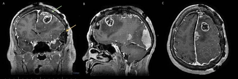
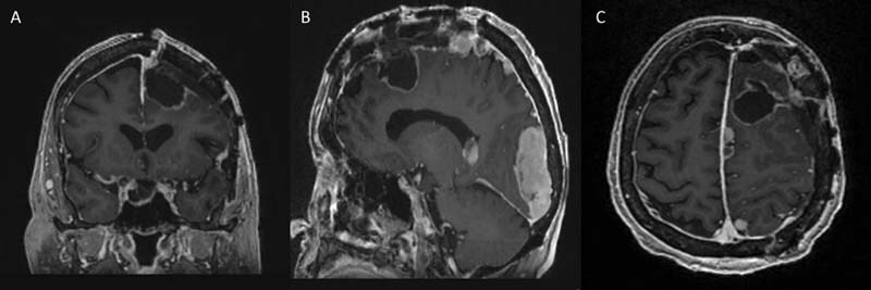
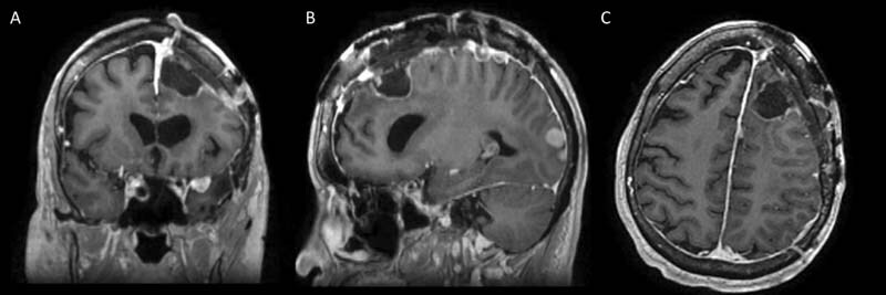
Introduction Collision tumors present as histologically different juxtaposed neoplasms within the same anatomical region, independent of the adjacent cell population. De novo intracranial collision tumors involving metachronous primary brain neoplasms alongside dural meningiomatosis are not well documented in the literature. Clinical Presentation We present staged surgical management of a 72-year-old female with known left hemispheric stable dural-based convexity mass lesions over 10 years and new-onset expressive aphasia and headaches. MRI had revealed left supratentorial dural-based enhanced masses consistent with en plaque meningiomatosis. Embolization angiography showed an unusual tumor blush from an aberrant branch of anterior cerebral artery suggesting a deeper focal intra-axial nature; a stage 1 craniotomy for dural-based tumor resection was completed with diagnosis of a meningioma (WHO grade 1). Intraoperatively, a distinct intra-axial deep discrete lesion was verified stereotactically, concordant with the location of tumor blush. The patient made a complete neurological recovery from a transient postoperative supplemental motor area syndrome in a week. Subsequent postoperative follow-up showed worsening of right hemiparesis and MRI showed an increase in residual lesion size and perilesional edema, which prompted a stage 2 radical resection of a glioblastoma, WHO grade 4. She improved neurologically after surgery with steroids and physical therapy. At 15 months following adjuvant therapy, she remains neurologically intact throughout the postoperative course, with no recurrent tumor on MRI. Conclusion A de novo glioblastoma presented as a masquerading lesion within hemispheric convexity meningiomatosis in an elderly patient with no prior radiation/phakomatosis, inciting a non-causal juxtapositional coexistence. The authors highlight rare pathognomonic angiographic findings and the rationale for two-staged resections of these collision lesions that led to excellent clinicoradiological outcome.

Keywords: case report; collision tumors; glioblastoma; glioma; meningioma; meningiomatosis; supplementary motor area syndrome.


Conflict of interest statement

Conflict of Interest None declared.

Figures

Fig. 1
Fig. 1
( A ) Axial section MRI brain with contrast depicting enhanced left-sided hemispheric dural meningiomatosis. ( B ) Coronal section showing a heterogeneous component of the “collision” tumor in deep left frontoparietal brain with perilesional edema and an extra-axial separate lesion in the Broca's area based on convexity dura mater. ( C ) Sagittal view showing a deep heterogeneous lesion with edema ( red arrow ), juxtaposed to dural meningiomatosis. ( D ) Better delineation of the heterogeneous component (glial) which was suspicious of glial origin, showing a discrete T2 FLAIR appearance compared with juxtaposed meningioma ( red arrow ) ( E ).
Fig. 2
Fig. 2
( A ) Left internal carotid artery injection showing early A3 branch supplying the superior pole of the likely intra-axial mass ( red arrow ). ( B ) Late arterial phase of the same ( red arrow ). ( C ) Contralateral injection from right internal carotid artery showing the relatively avascular parasagittal lesion and independent supply of the left-sided deep lesion ( red arrow ). ( D ) Lateral view showing relatively avascular parasagittal meningiomatosis ( see green arrows ) and left external carotid arteriogram showing a tumor blush at the frontotemporal extra-axial mass based off the dura mater ( red arrow ) ( E ).
Fig. 3
Fig. 3
( A ) Microscopic sections show a moderately cellular lesion with a whorling and syncytial growth pattern. ( B ) The tumor cells show round, uniform nuclei and eosinophilic cytoplasm. Abundant psammomatous calcifications are present. ( C ) Biopsy of the deeper suspicious focus identified on angiography shows an infiltrating glioma with high cellularity. ( D,E ) Higher power images display pleomorphic nuclei, brisk mitotic figures, prominent microvascular proliferation and pseudopalisading necrosis, characteristic of glioblastoma (WHO grade 4). ( F ) IDH-1 was negative on immunohistochemical analysis, showing a wild-type profile.
Fig. 4
Fig. 4
( A ) Coronal cranial MRI with contrast showing a heterogeneous enhancing frontoparietal intra-axial tumor ( green arrow ) likely of glial origin with perilesional white matter edema and postoperative changes of focal meningiomatosis resection and Simpson grade 1 resection of lesion near the Broca's area ( yellow arrow ). ( B ) Post stage 1 craniotomy, sagittal contrast MRI brain showing the heterogeneous enhanced frontoparietal intra-axial tumor, which was masqueraded by juxtaposed meningiomas. ( C ) Axial cranial MRI view with contrast showing the frontoparietal intra-axial tumor with remarkable perilesional white matter edema and postoperative changes of focal meningiomatosis resection.
Fig. 5
Fig. 5
( A, B, C ) Early postoperative contrast MRI after stage 2 focal re-craniotomy for microsurgical radical resection of the intra-axial mass showing radical resection of the lesion. ( A ) coronal, ( B ) sagittal, and ( C ) axial sections
Fig. 6
Fig. 6
( A, B, C ) Six months follow-up of postoperative contrast MRI after stage 2 focal re-craniotomy for microsurgical radical resection of the intra-axial mass shows radical resection. ( A ) coronal, ( B ) sagittal, and ( C ) axial sections

References

    1. Zhao Y, Zhang H, Lian W. Collision tumors composed of meningioma and growth hormone-secreting pituitary adenoma in the sellar region: case reports and a literature review. Medicine (Baltimore) 2017;96(50):e9139. - PMC - PubMed
    1. Thon N, Kreth S, Kreth F-W. Personalized treatment strategies in glioblastoma: MGMT promoter methylation status. OncoTargets Ther. 2013;6:1363–1372. - PMC - PubMed
    1. Sung C T, Shetty A, Menias C O. Collision and composite tumors; radiologic and pathologic correlation. Abdom Radiol (NY) 2017;42(12):2909–2926. - PubMed
    1. Ohgaki H, Kleihues P. Genetic pathways to primary and secondary glioblastoma. Am J Pathol. 2007;170(05):1445–1453. - PMC - PubMed
    1. Lee H S, Kim J H, Lee J-I. Glioblastoma following radiosurgery for meningioma. J Korean Neurosurg Soc. 2012;51(02):98–101. - PMC - PubMed

LinkOut - more resources