. 2022 Mar 23;4(3):000324.
doi: 10.1099/acmi.0.000324. eCollection 2022.

Cholera outbreak: antibiofilm activity, profiling of antibiotic-resistant genes and virulence factors of toxigenic Vibrio cholerae isolates reveals concerning traits

Affiliations

Cholera outbreak: antibiofilm activity, profiling of antibiotic-resistant genes and virulence factors of toxigenic Vibrio cholerae isolates reveals concerning traits

Silas O Awuor et al. Access Microbiol. .

Abstract

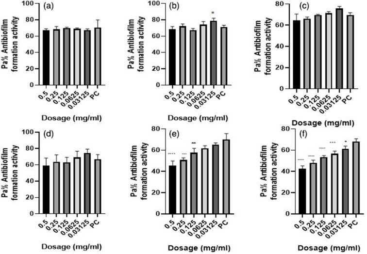
Vibrio cholerae is a biofilm-forming pathogen with various virulence phenotypes and antimicrobial resistance traits. Phenotypic characteristics play a critical role in disease transmission and pathogenesis. The current study elucidated antibiofilm formation activity, profiled antibiotic-resistant genes and virulence factors of toxigenic Vibrio cholerae isolates from the cholera outbreak in Kisumu County, Kenya. Vibrio cholerae O1 isolates collected during the 2017 cholera outbreak in Kisumu County, Kenya, were utilized. Biofilm and virulence factors were profiled using standard procedures. The study confirmed 100 isolates as Vibrio cholerae , with 81 of them possessing cholera toxin gene (ctxA). Additionally, 99 of the isolates harboured the toxR gene. The study further revealed that 81 and 94 of the isolates harboured the class I integron (encoded by inDS gene) and integrating conjugative element (ICE), respectively. Antibiotic resistance assays confirmed tetracycline resistance genes as the most abundant (97 isolates). Among them were seven isolates resistant to commonly used antibiotics. The study further screened the isolates for antibiofilm formation using various antibiotics. Unlike the four strains (03/17-16, 02/17-09, 04/17-13), three of the strains (04/17-07, 06/17-14 and 05/17-03) did not form biofilms. Further, all the seven isolates that exhibited extensive antibiotic resistance produced haemolysin while 71.42%, 85.71 and 71.42 % of them produced protease, phospholipases and lipase, respectively. This study provides and in-depth understanding of essential features that were possibly responsible for V. cholerae outbreak. Understanding of these features is critical in the development of strategies to combat future outbreaks.

Keywords: Vibrio cholerae; antibiofilm activity; antibiotics; resistant genes profiling; virulence factors.


Conflict of interest statement

The authors declare that there are no conflicts of interest.

Figures

Fig. 1.
Fig. 1.
Gel electrophoresis for confirmation of various genes extracted from the seven isolates of V. cholerae that were resistant to various commonly used antibiotics with plate A representing the ctxA gene in clinical V. cholerae , plate B representing inDS, toxR and int genes in clinical V. cholerae isolate and plate C representing tetA and Ery genes in clinical V. cholerae isolates. The legends: lane 1 (05/17–03 isolate*); lane 2 (06/17–14 isolate*); lane 3 (03/17–16 isolate*); lane 4 (04/17–07 isolate*); lane 5 (02/17–09 isolate*); lane 6 (04/17-13Isolate*) and lane 7 (06/17–07 isolate*). Lane M1 for plate A represents 2 kb DNA Size Marker- Hyper ladder I ctxA band 289 bp; lane M1 for plate B represents 2 kb DNA Size Marker- Hyper ladder I inDS 869 bp, toxR 779 bp and int 481 bp and lane M1 for plate C represents 2 kb DNA Size Marker- Hyper ladder I, tetA 862 bp and Ery 564 bp. * The figures in brackets represent strain numbers.
Fig. 2.
Fig. 2.
Antibiofilm formation activity against isolate 03/17–16 of V. cholerae against various antibiotics: (a) tetracycline (b) ampicillin, (c) amoxicillin, (d) cotrimoxazole, (e) erythromycin and (f) nalidixic acid; PC=P. aeruginosa – Positive control (n=3, ANOVA Dunnett’s multiple comparisons test; *P=0.05; **P=0.01; ***P=0.001; ****P=0.0001).
Fig. 3.
Fig. 3.
Antibiofilm formation activity against isolate 02/17–09 of V. cholerae against various antibiotics: (a) tetracycline, (b) ampicillin, (c) amoxicillin, (d) cotrimoxazole, (e) erythromycin and (f) nalidixic acid; PC=P. aeruginosa – positive control (n=3, Anova Dunnett’s multiple comparisons test; *P=0.05; **P=0.01; ***P=0.001; ****P=0.0001).
Fig. 4.
Fig. 4.
Antibiofilm formation activity against isolate 04/17–13 of V. cholerae against various antibiotics: (a) tetracycline (b) ampicillin, (c) amoxicillin, (d) cotrimoxazole, (e) erythromycin and (f) nalidixic acid; PC=P. aeruginosa – positive control (n=3, Anova Dunnett’s multiple comparisons test; *P=0.05; **P=0.01; ***P=0.001; ****P=0.0001). Against this test isolate 04/17–13, inhibitory activities were observed to be more at lower dosages than at high dosages.
Fig. 5.
Fig. 5.
Antibiofilm formation activity against isolate 05/17–07 of V. cholerae against various antibiotics: (a) tetracycline (b) ampicillin, (c) amoxicillin, (d) cotrimoxazole, (e) erythromycin and (f) nalidixic Acid; PC=P. aeruginosa – positive control (n=3, Anova Dunnett’s multiple comparisons test; *P=0.05; **P=0.01; ***P=0.001; ****P=0.0001).
Fig. 6.
Fig. 6.
Antibiofilm formation activity against isolate 05/17–03 of V. cholerae against various antibiotics: (a) tetracycline, (b) ampicillin, (c) amoxicillin, (d) cotrimoxazole, (e) erythromycin and (f) nalidixic acid; PC=P. aeruginosa – positive control (n=3, Anova Dunnett’s multiple comparisons test; *P=0.05; **P=0.01; ***P=0.001; ****P=0.0001).
Fig. 7.
Fig. 7.
Antibiofilm formation activity against isolate 06/17–14 of V. cholerae against various antibiotics: (a) tetracycline, (b) ampicillin, (c) amoxicillin, (d) cotrimoxazole, (e) erythromycin and (f) nalidixic acid; PC=P. aeruginosa – positive control (n=3, Anova Dunnett’s multiple comparisons test; *P=0.05; **P=0.01; ***P=0.001; ****P=0.0001).
Fig. 8.
Fig. 8.
Showing the distribution of the seven isolates verses the various virulence traits they produced.
Fig. 9.
Fig. 9.
Representation of the various virulence traits produced by the V. cholerae isolates (a). The distribution of Protease enzyme production of V. cholerae (b). The Phospholipase enzyme production of V. cholerae (c). The haemolysin enzyme production of V. cholerae – haemolytic activity (d). Lipase enzyme production of V. cholerae .

References

    1. Halpern M, Izhaki I. Fish as hosts of Vibrio cholerae . Front Microbiol. 2017;8:282. doi: 10.3389/fmicb.2017.00282. - DOI - PMC - PubMed
    1. Sakib SN, Reddi G, Almagro-Moreno S, DiRita VJ. Vibrio JB special issue. minireview environmental role of pathogenic traits in Vibrio cholerae . J Bacteriol. 2018;200:17. doi: 10.1128/JB.00795-17. - DOI - PMC - PubMed
    1. Matthey N, Blokesch M. The DNA-Uptake process of naturally competent Vibrio cholerae . Trends Microbiol. 2016;24:98–110. doi: 10.1016/j.tim.2015.10.008. - DOI - PubMed
    1. Cairncross S, Feachem R. Environmental Health Engineering in the Tropics. Vol. 134. Routledge; 2018. Water, sanitation and disease control; pp. 865–871. vol. - DOI
    1. Craig RK. Cholera and climate change: pursuing public health adaptation strategies in the face of scientific debate. Houst J Health Law Policy. 2018;3:964–999. doi: 10.1015/S22211691(14)60185-9. - DOI

LinkOut - more resources