

Spontaneously Arrested Bilateral Primary Congenital Glaucoma: A Case Report From Ethiopia
- PMID: 35693561
- PMCID: PMC9175216
- DOI: 10.4314/ejhs.v32i2.27
Spontaneously Arrested Bilateral Primary Congenital Glaucoma: A Case Report From Ethiopia
Abstract
Background: Primary congenital glaucoma is potentially blinding condition characterized by elevated intraocular pressure and optic disc cupping. It is typically bilateral and usually manifest in the first year of life. Spontaneously arrested primary congenital glaucoma can occur, but it is very rare.
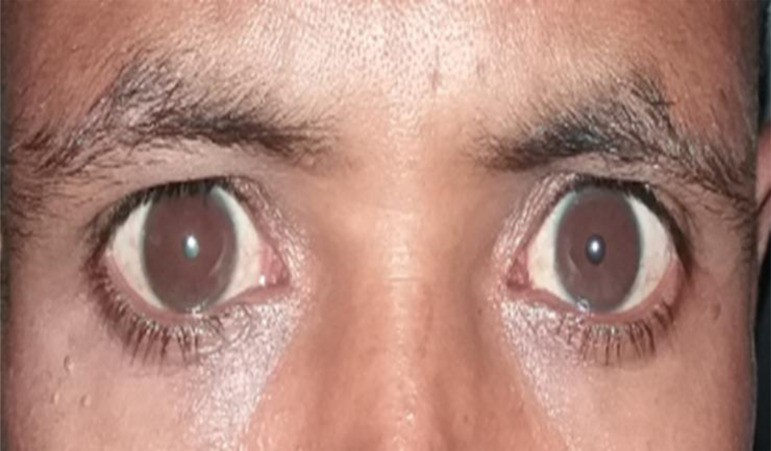
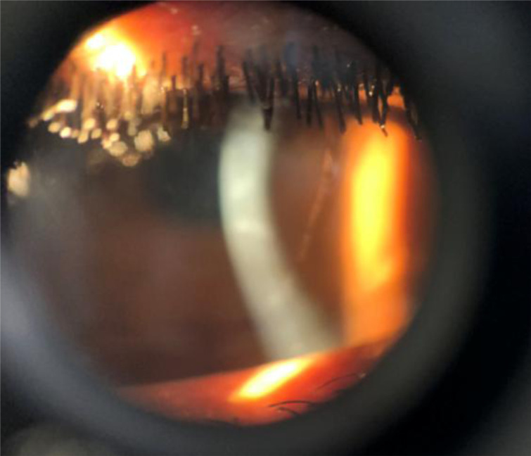
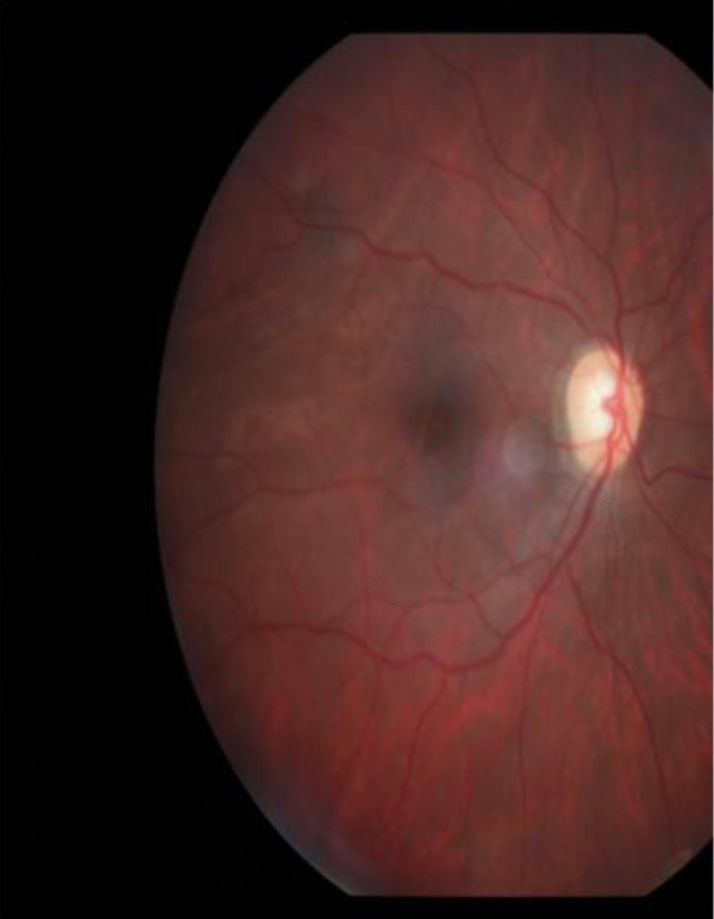
Case report: A 32-year-old male patient from North Shewa presented to the department of ophthalmology, Menelik II Hospital with deterioration of vision. On examination he had large corneas with horizontal diameter of 14 mm, increased axial length, faint corneal stromal opacity and Haab's striae of both eyes. Anterior chamber angles were wide open. His intraocular pressure, optic nerve head appearance and visual field in both eyes were normal. He had subluxated dense cataract of the right eye.
Conclusion: Late presentation with sequelae of primary congenital glaucoma without optic neuropathy is possible. Regular follow-up of spontaneously arrested congenital glaucoma and scleral fixation of intraocular lens is recommended.
Keywords: Megalocornea; Primary congenital glaucoma; Subluxated cataract; spontaneously arrested primary congenital glaucoma.
© 2022 Kalkidan S.Alayu, et al.
Figures
Similar articles
-
Premature Expression of Pseudoexfoliation Syndrome With Presenile Cataract in a 28-Year-Old Lady.J Glaucoma. 2019 Jul;28(7):e115-e117. doi: 10.1097/IJG.0000000000001219. J Glaucoma. 2019. PMID: 30807439
-
A case of primary congenital glaucoma: a diagnostic dilemma.Optometry. 2007 Apr;78(4):167-75. doi: 10.1016/j.optm.2006.10.016. Optometry. 2007. PMID: 17400138
-
Assessment of diagnostic criteria in management of infantile glaucoma. An analysis of tonometry, optic disc cup, corneal diameter and axial length.Int Ophthalmol. 1996-1997;20(1-3):21-7. doi: 10.1007/BF00212940. Int Ophthalmol. 1996. PMID: 9112158
-
Phacolytic glaucoma: A nearly forgotten entity.Eur J Ophthalmol. 2020 Sep;30(5):NP32-NP35. doi: 10.1177/1120672119841972. Epub 2019 Apr 5. Eur J Ophthalmol. 2020. PMID: 30950286 Review.
-
[Early primary congenital glaucoma].Oftalmologia. 2004;48(2):15-21. Oftalmologia. 2004. PMID: 15341093 Review. Romanian.
Cited by
-
Intrinsic disorder in CYP1B1 and its implications in primary congenital glaucoma pathogenesis.J Proteins Proteom. 2025 May 13:10.1007/s42485-025-00186-8. doi: 10.1007/s42485-025-00186-8. Online ahead of print. J Proteins Proteom. 2025. PMID: 40821877 Free PMC article.
References
-
- Beck AD, Chang TCP, Freedman SF. Definition, Classification, Differential Diagnosis. In: Weinreb RN, et al., editors. Childhood Glaucoma: Consensus Series 9. Amsterdam: Kugler; 2013.
-
- Beck Allen, Chang Ta Chen Peter. Glaucoma: Definitions and Classification. [Aug 18, 2016];Am J Opthalmol. 2016 170:214–222. Available from www.aao.org/disease-review/glaucoma.
-
- Chang L, et al. A review of medical treatment of pediatric glaucoma at Moorfield's Eye Hospital. J Glauoma. 2013;22(8):601–607. - PubMed
Publication types
MeSH terms
LinkOut - more resources
Full Text Sources
Medical