. 2022 May;14(5):1663-1673.
doi: 10.21037/jtd-22-20.

Study design for a multicenter, randomized controlled trial evaluating the diagnostic value of ultrathin bronchoscope compared to thin bronchoscope without fluoroscopy for peripheral pulmonary lesions

Affiliations

Study design for a multicenter, randomized controlled trial evaluating the diagnostic value of ultrathin bronchoscope compared to thin bronchoscope without fluoroscopy for peripheral pulmonary lesions

Xintong Feng et al. J Thorac Dis. 2022 May.

Abstract

Background: Ultrathin bronchoscope (UTB) with a 3.0-mm outer diameter and a 1.7-mm working channel currently appeared as a potential tool for better biopsy and diagnosis of peripheral pulmonary lesions (PPLs) by accessing more distal bronchus. However, published research is primarily limited to diagnosis value of UTB for PPLs with fluoroscopy, the value of UTB compared with thin bronchoscope (TB) without fluoroscopy guidance has not been determined yet.

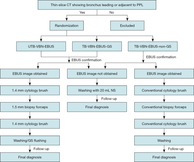
Methods: We design a prospective, randomized, controlled, non-inferior, multicenter study aiming to evaluate the diagnostic value and safety of UTB for PPLs with the guidance of virtual bronchoscopic navigation (VBN) combined with endobronchial ultrasound (EBUS) without fluoroscopy by comparing to TB. The study aims to enroll 578 patients presenting for evaluation of PPLs at five clinical sites in China. Subjects will be randomized to UTB-VBN-EBUS group, TB-VBN-EBUS-guide sheath (GS) group, and TB-VBN-EBUS-non-GS group. Primary endpoint is the diagnostic yield of PPLs. The total examination time, duration of finding lesions, the proportion of lesions visible by radial EBUS, factors affecting the diagnostic yield, difference in the bronchus level reached with the bronchoscope, difference in diagnostic yield, and complication rate will be determined as secondary endpoints. The primary endpoint will be followed-up at least 6-month post-procedure and 1-month post-procedure for safety endpoint.

Discussion: Study enrollment began in March 2021. Our preliminary experience reveals that UTB is a powerful tool in the diagnosis of PPLs even without fluoroscopy. The results of the current study will compensate the limitations of the previous research, further provide evidence of UTB in diagnosing PPLs without fluoroscopy.

Trial registration: ClinicalTrials.gov NCT04571476. Registered on 30 September 2020.

Keywords: Ultrathin bronchoscope (UTB); endobronchial ultrasound (EBUS); peripheral pulmonary lesions (PPLs); thin bronchoscope (TB); virtual bronchoscopic navigation (VBN).


Conflict of interest statement

Conflicts of Interest: All authors have completed the ICMJE uniform disclosure form (available at https://jtd.amegroups.com/article/view/10.21037/jtd-22-20/coif). All authors report funding from Olympus (Beijing) sales and service Co., Ltd.

Figures

Figure 1
Figure 1
Flow chart of UTB and TB for the diagnosis of PPL. Patients with PPLs who meet the criteria will be randomized into UTB-VBN-EBUS group, TB-VBN-EBUS-GS group, and TB-VBN-EBUS-non-GS group. EBUS will be used to verify the location of the lesion. Sampling will be performed following the sequence of brushing, biopsy, brushing, washing or GS flushing in lesions with EBUS image obtained. Washing with 20 mL NS will be performed as a supplementary procedure in lesions that can’t obtain EBUS image. All patients will be followed-up for at least 6 months post-procedure for the final diagnosis. CT, computed tomography; PPL, peripheral pulmonary lesion; UTB, ultrathin bronchoscope; VBN, virtual bronchoscopic navigation; EBUS, endobronchial ultrasound; TB, thin bronchoscope; GS, guide sheath; NS, normal saline.
Figure 2
Figure 2
UTB and other instruments used in the study. (A) UTB (BF-MP290F, Olympus). (B) a, UTB (BF-MP290F) with a 3.0-mm outer diameter and a 1.7-mm working channel; b, TB (BF-P290) with a 4.2-mm outer diameter and a 2.0-mm working channel; c, radial-type probe EBUS with 1.4-mm outer diameter (UM-S20-17S); d, GS with 1.95-mm outer diameter; e, 1.5-mm biopsy forceps; f, 1.8-mm conventional biopsy forceps; g, 1.4-mm cytology brush; h, 1.8-mm conventional cytology brush. UTB, ultrathin bronchoscope; TB, thin bronchoscope; EBUS, endobronchial ultrasound; GS, guide sheath.
Figure 3
Figure 3
Instruments used in the three groups. (A) UTB-VBN-EBUS group: UTB (BF-MP290F), 1.4-mm radial-type probe EBUS (UM-S20-17S), 1.5-mm biopsy forceps, 1.4-mm cytology brush, from left to right. (B) TB-VBN-EBUS-GS group: TB (BF-P290), 1.95-mm GS, 1.4-mm radial-type probe EBUS (UM-S20-17S), 1.5-mm biopsy forceps, 1.4-mm cytology brush, from left to right. (C) TB (BF-P290), 1.4-mm radial-type probe EBUS (UM-S20-17S), 1.8-mm conventional biopsy forceps, 1.8-mm conventional cytology brush, from left to right. UTB, ultrathin bronchoscope; VBN, virtual bronchoscopic navigation; EBUS, endobronchial ultrasound; TB, thin bronchoscope; GS, guide sheath.
Figure 4
Figure 4
A representative case of UTB used to diagnose PPL. A 66-year-old male patient with a small solid peripheral pulmonary nodule underwent TBLB using UTB with the guidance of VBN combined with EBUS without fluoroscopy. The pathology showed malignant tumor cells. (A) Chest CT presented a small pulmonary nodule (red arrow) measuring 19.0×18.2×21.3 mm with lobulation and spiculate in the right upper lobe. (B) The virtual bronchial tree with the target lesion (the red spot pointed by red arrow) and the navigation pathway (along with the blue line) planned by VBN system. (C) The virtual bronchoscopic view with red spot highlighted the target lesion and the red arrow pointed the bronchus leading to it. (D) Ultrathin bronchoscopic view of target bronchus (red arrow) leading to the lesion. (E) EBUS image of the lesion. (F) ROSE of the biopsy specimens by Diff-Quik staining (×400). (G) The H&E staining histopathology of the biopsy specimens (×400). Scale bars: 20 µm. This image is published with the patient’s consent. UTB, ultrathin bronchoscope; PPL, peripheral pulmonary lesion; TBLB, transbronchial lung biopsy; VBN, virtual bronchoscopic navigation; EBUS, endobronchial ultrasound; CT, computed tomography; ROSE, rapid on-site evaluation; H&E, hematoxylin and eosin.

Similar articles

Cited by

References

    1. Bray F, Ferlay J, Soerjomataram I, et al. Global cancer statistics 2018: GLOBOCAN estimates of incidence and mortality worldwide for 36 cancers in 185 countries. CA Cancer J Clin 2018;68:394-424. 10.3322/caac.21492 - DOI - PubMed
    1. National Lung Screening Trial Research Team , Aberle DR, Adams AM, et al. Reduced lung-cancer mortality with low-dose computed tomographic screening. N Engl J Med 2011;365:395-409. 10.1056/NEJMoa1102873 - DOI - PMC - PubMed
    1. Tomiyama N, Yasuhara Y, Nakajima Y, et al. CT-guided needle biopsy of lung lesions: a survey of severe complication based on 9783 biopsies in Japan. Eur J Radiol 2006;59:60-4. 10.1016/j.ejrad.2006.02.001 - DOI - PubMed
    1. Schreiner AM, Jones JG, Swistel AJ, et al. Transthoracic fine needle aspiration resulting in implantation metastasis in the superficial tissues of the breast. Cytopathology 2013;24:58-60. 10.1111/j.1365-2303.2012.00977.x - DOI - PubMed
    1. Stringfield JT, Markowitz DJ, Bentz RR, et al. The effect of tumor size and location on diagnosis by fiberoptic bronchoscopy. Chest 1977;72:474-6. 10.1378/chest.72.4.474 - DOI - PubMed

Associated data