. 2022 Jun 1:72:311-370.
doi: 10.3897/vz.72.e84732.

Cranial morphology of the orectolobiform shark, Chiloscyllium punctatum Müller & Henle, 1838

Affiliations

Cranial morphology of the orectolobiform shark, Chiloscyllium punctatum Müller & Henle, 1838

Manuel Andreas Staggl et al. Vertebr Zool. .

Abstract

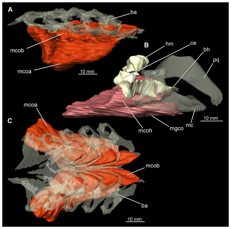
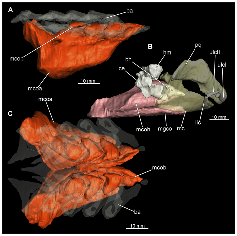
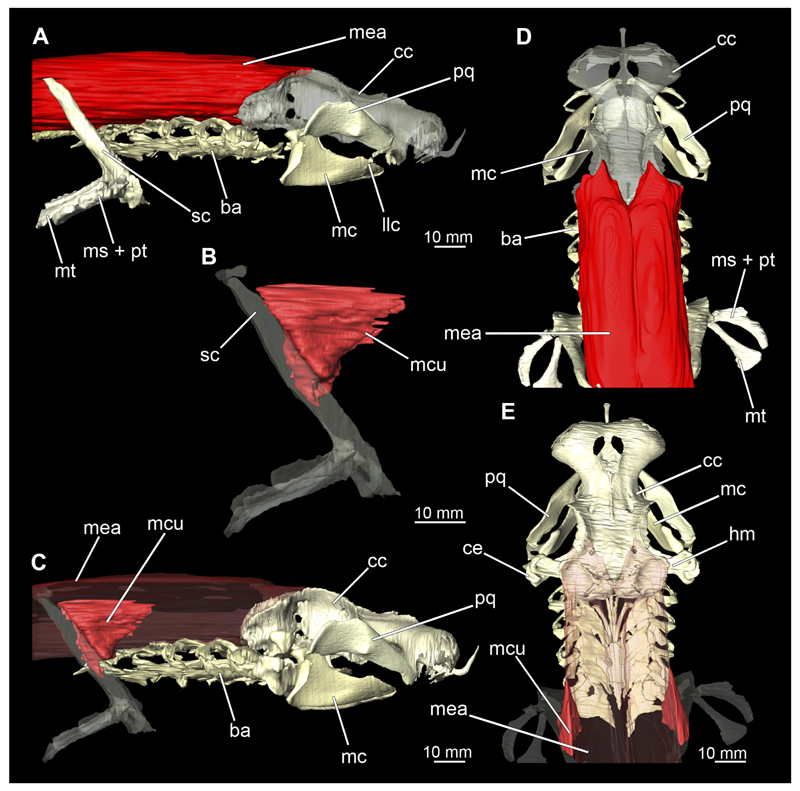
Elasmobranchs, comprising sharks, skates, and rays, have a long evolutionary history extending back into the Palaeozoic. They are characterized by various unique traits including a predominantly cartilaginous skeleton, superficial prismatic phosphatic layer, and permanent tooth replacement. Moreover, they exhibit a more or less marked sexual dimorphism. Especially the morphology of the chondrocranium and the elements of the whole cranial region of extant and extinct chondrichthyans can provide valuable information about corresponding functions, e.g. the feeding apparatus might reflect the diet of the animals. However, studies on sexual dimorphisms are lacking in orectolobiform sharks, therefore, little is known about possible sexual dimorphic characters in the cranial region in this group. For this reason, we present in this study a comprehensive morphological description of the cranial region of the brownbanded bamboo shark Chiloscyllium punctatum Müller & Henle, 1838, with a special focus on its sexual dimorphic characters. Our results reveal clear morphological differences in both sexes of the examined C. punctatum specimens, particularly in the chondrocranium and the mandibular arch. The female specimen shows a comparatively more robust and compact morphology of the chondrocranium. This pattern is also evident in the mandibular arch, especially in the palatoquadrate. The present study is the first to describe the morphology of an orectolobiform shark species in detail using both manual dissection and micro-CT data. The resulting data furthermore provide a starting point for pending studies and are intended to be a first step in a series of comparative studies on the morphology of the cranial region of orectolobiform sharks, including the determination of possible sexual dimorphic characteristics.

Keywords: Chiloscyllidae; Chondrichthyes; Galeomorphii; dissection; micro-CT.


Conflict of interest statement

The authors declare no conflict of interest

Figures

Figure 1
Figure 1. Chiloscyllium punctatum, male: Different perspectives of the anterior part of the body up to the pelvic girdle of the male specimen.
A dorsal, B lateral and C ventral views. Abbreviations: ba, barbell; cl, cloaca; dol, dorsal fin; ey, eye; go, gill opening; inc, incision; mo, mouth nopening; no, nostril; pec, pectoral fin; pel, pelvic fin; sp, spiracle.
Figure 2
Figure 2. Chiloscyllium punctatum, female: Different perspectives of the anterior part of the body up to the pelvic girdle of the female specimen.
A dorsal, B lateral and C ventral views. Abbreviations: ba, barbell; cl, cloaca; dol, dorsal fin; ey, eye; go, gill opening; inc, incision; mo, mouth nopening; no, nostril; pec, pectoral fin; pel, pelvic fin; sp, spiracle.
Figure 3
Figure 3. Chiloscyllium punctatum, male: Different perspectives of the anterior part of dissected specimen. In this dissection step, only the skin was removed. Large parts of the connective tissue were left on the object. The subcutaneous nerve cords and the ampullae of Lorenzini are well visible.
A dorsal, B lateral, C ventral side of the rostrum and D ventral views. Abbreviations: ba, barbell; dal, dorsal ampullae of Lorenzini; ey, eye; lal, lateral ampullae of Lorenzini; mo, mouth opening; nc, nasal capsule; no, nostril; pec, pectoral fin; sp, spiracle; val, ventral ampullae of Lorenzini.
Figure 4
Figure 4. Chiloscyllium punctatum, female: Different perspectives of the anterior part of dissected specimen. In this dissection step, only the skin was removed. Large parts of the connective tissue were left on the object. The subcutaneous nerve cords and the ampullae of Lorenzini are well visible. The weak condition of the soft tissues is also evident.
A dorsal, B lateral, C ventral views. Abbreviations: ba, barbell; dal, dorsal ampullae of Lorenzini; ey, eye; lal, lateral ampullae of Lorenzini; mo, mouth opening; nc, nasal capsule; no, nostril; pec, pectoral fin; sp, spiracle; val, ventral ampullae of Lorenzini.
Figure 5
Figure 5. Chiloscyllium punctatum, male: Different views of the chondocranium of the specimen.
A lateral, B posterior, C anterior, D ventral and E dorsal views as rendered surface. Abbreviations: aop, Antorbital process; elf, endolymphatic foramen;.fm, foramen magnum; fonv, foramen of orbito-nasal vein; foa, foramen for orbital artery; fII, foramen of cranial nerv II; fV, foramen of cranial nerv V; fVprof, foramen of profundus branch of cranial nerv V; fVsup, foramen of superficial branch of cranial nerv V; fVII, foramen of cranial nerv VII; fIX, foramen of cranial nerv IX; fX, foramen of cranial nerv X; ins, intranasal septum; nc, nasal capsule; ob, orbita; pba, foramen for pseudobranchial artery; pbr, Palatobasal ridge; pff, prefrontal fontanelle; pit, foramen for pituitary vein; plf, perilympatic foramen; pop, postorbital process; rp, rostral process; soc, supraorbital crest; sof, supraorbital foramen; spr, sphenopterotic ridge.
Figure 6
Figure 6. Chiloscyllium punctatum, male: Different views of the chondocranium of the dissected specimen.
A lateral, B posterior, C anterior, D ventral and E dorsal views as rendered surface. Abbreviations: aop, Antorbital process; elf, endolymphatic foramen;.fm, foramen magnum; fonv, foramen of orbito-nasal vein; fII, foramen of cranial nerv II; fV, foramen of cranial nerv V; fVprof, foramen of profundus branch of cranial nerv V; fVsup, foramen of superficial branch of cranial nerv V; fVII, foramen of cranial nerv VII; fIX, foramen of cranial nerv IX; fX, foramen of cranial nerv X; ins, intranasal septum; nc, nasal capsule; ob, orbita; pba, foramen for pseudobranchial artery; pbr, Palatobasal ridge; pff, prefrontal fontanelle; pit, foramen for pituitary vein; plf, perilympatic foramen; pop, postorbital process; rp, rostral process; soc, supraorbital crest; sof, supraorbital foramen; spr, sphenopterotic ridge.
Figure 7
Figure 7. Chiloscyllium punctatum, female: Different views of the chondocranium of the specimen.
A lateral, B posterior, C anterior, D ventral and E dorsal views as rendered surface. Abbreviations: aop, Antorbital process; elf, endolymphatic foramen; fm, foramen magnum; fonv, foramen of orbito-nasal vein; foa, foramen for orbital artery; fII, foramen of cranial nerv II; fV, foramen of cranial nerv V; fVprof, foramen of profundus branch of cranial nerv V; fVsup, foramen of superficial branch of cranial nerv V; fVII, foramen of cranial nerv VII; fIX, foramen of cranial nerv IX; fX, foramen of cranial nerv X; ins, intranasal septum; nc, nasal capsule; ob, orbita; pba, foramen for pseudobranchial artery; pbr, Palatobasal ridge; pff, prefrontal fontanelle; pit, foramen for pituitary vein; plf, perilympatic foramen; pop, postorbital process; rp, rostral process; soc, supraorbital crest; sof, supraorbital foramen; spr, sphenopterotic ridge.
Figure 8
Figure 8. Chiloscyllium punctatum, female: Different views of the chondocranium of the dissected specimen.
A lateral, B posterior, C anterior, D ventral and E dorsal views as rendered surface. Abbreviations: aop, Antorbital process; elf, endolymphatic foramen;. fm, foramen magnum; fonv, foramen of orbito-nasal vein; fII, foramen of cranial nerv II; fV, foramen of cranial nerv V; fVprof, foramen of profundus branch of cranial nerv V; fVsup, foramen of superficial branch of cranial nerv V; fVII, foramen of cranial nerv VII; fIX, foramen of cranial nerv IX; fX, foramen of cranial nerv X; ins, intranasal septum; nc, nasal capsule; ob, orbita; pba, foramen for pseudobranchial artery; pbr, Palatobasal ridge; pff, prefrontal fontanelle; pit, foramen for pituitary vein; plf, perilympatic foramen; pop, postorbital process; rp, rostral process; soc, supraorbital crest; sof, supraorbital foramen; spr, sphenopterotic ridge; V, Nervus trigeminalis (CN V).
Figure 9
Figure 9. Chiloscyllium punctatum, male: Olphactory sacs within the nasal capsules.
A dorsal, B lateral, C ventral, arrows indicating direction of waterflow, D olphactory sacs anterior, arrows indicating direction of waterflow, E olphactory sacs ventral, arrows indicating direction of waterflow, F olphactory sacs within chondrocranium anterior and G transversal cross section through rostral area of the specimen, yellow markings showing the epithelium, arrows pointing to the folding of the tissue. Abbreviations: al, ampullae of Lorenzini; cc, chondrocranium; ins, intranasal septum; nc, nasal capsules; os, ophactory sacs, rp, rostral process.
Figure 10
Figure 10. Chiloscyllium punctatum, female: Olphactory sacs within the nasal capsules.
A dorsal, B lateral, C ventral, arrows indicating direction of waterflow, D olphactory sacs anterior, arrows indicating direction of waterflow, E olphactory sacs ventral, arrows indicating direction of waterflow, F olphactory sacs within chondrocranium anterior and G transversal cross section through rostral area of the specimen, yellow markings showing the epithelium, arrows pointing to the folding of the tissue. Abbreviations: al, ampullae of Lorenzini; cc, chondrocranium; ins, intranasal septum; nc, nasal capsules; os, ophactory sacs, rp, rostral process.
Figure 11
Figure 11. Chiloscyllium punctatum, male: Different views of the inner ear of the specimen.
A dorsal view within the chondro-cranium, B lateral view within the chondrocranium, C posterior view and D lateral view as rendered surface. Abbreviations: amp, Ampulla; asd, anterior semicircular canal, cc, chondrocranium; ed, endolymphatic duct; hsd, horizontal semicircular canal, psd, posterior semicircular canal, rp, rostral process; sa, sacculus.
Figure 12
Figure 12. Chiloscyllium punctatum, female: Different views of the inner ear of the specimen.
A dorsal view within the chondro-cranium, B lateral view within the chondrocranium, C posterior view and D lateral view as rendered surface. Abbreviations: amp, Ampulla; asd, anterior semicircular canal, cc, chondrocranium; ed, endolymphatic duct; hsd, horizontal semicircular canal, psd, posterior semicircular canal, rp, rostral process; sa, sacculus.
Figure 13
Figure 13. Chiloscyllium punctatum, male: Different views of the mandibular arch of the specimen.
A anterior, B posterior and C lateral views as rendered surface. Abbreviations: amp, adductor mandibular process; apq, ascending process of the palatoquadratum; fo, foramen; mc, Meckel’s cartilage; mk, mandibular knob; pq, palatoquadratum; qjl, lateral quadratomandibular joint; qjm, medial quadratomandibular joint; sust, sustentaculum; sym, symphysis.
Figure 14
Figure 14. Chiloscyllium punctatum, male: Different views of the mandibular and the hyoid arch of the dissected specimen.
A anterior, B posterior, C dorsal, D ventral and E lateral views of the first and second arch of the splanchiocranium, F lower teeth, and G upper and lower teeth. Abbreviations: amp, adductor mandibular process; apq, ascending process of the palatoquadratum; bh, basihyale; ce, ceratohyal; fo, foramen; hm, hyomandibula; lt, lower teeth; mc, Meckel’s cartilage; mk, mandibular knob; pq, palatoquadratum; qjl, lateral quadratomandibular joint; qjm, medial quadratomandibular joint; sust, sustentaculum; sym, symphysis; ut, upper teeth.
Figure 15
Figure 15. Chiloscyllium punctatum, female: Different views of the mandibular arch of the specimen.
A anterior, B posterior and C lateral views as rendered surface. Abbreviations: amp, adductor mandibular process; apq, ascending process of the palatoquadratum; fo, foramen; mc, Meckel’s cartilage; mk, mandibular knob; pq, palatoquadratum; qjl, lateral quadratomandibular joint; qjm, medial quadratomandibular joint: sust, sustentaculum; sym, symphysis;
Figure 16
Figure 16. Chiloscyllium punctatum, female: Different views of the mandibular and the hyoid arch of the dissected specimen.
A anterior, B lateral, C dorsal and D ventral views of the first and second arch of the splanchiocranium, E lower teeth lateral (arrows pointing to the cusplets), F lower teeth anteriat (arrows pointing to the cusplets) and G upper teeth anteriat. Abbreviations: amp, adductor mandibular process; apq, ascending process of the palatoquadratum; bh, basihyale; ce, ceratohyal; fo, foramen; hm, hyomandibula; lt, lower teeth; mc, Meckel’s cartilage; mk, mandibular knob; pq, palatoquadratum; qjl, lateral quadratomandibular joint; qjm, medial quadratomandibular joint; sust, sustentaculum; sym, symphysis; ut, upper teeth.
Figure 17
Figure 17. Chiloscyllium punctatum, male: Different views of the labial cartilage attached to the mandibular arch of the specimen.
A anterior, B lateral and C ventral views as rendered surface. Abbreviations: cc, chondrocranium; fo, foramen; llc, lower labial cartilage; mc, Meckel’s cartilage; pq, palatoquadratum; ulcI, upper labial cartilage I; ulcII, upper labial cartilage II.
Figure 18
Figure 18. Chiloscyllium punctatum, female: Different views of the labial cartilage attached to the mandibular arch of the specimen.
A anterior, B lateral and C ventral views as rendered surface. Abbreviations: cc, chondrocranium; fo, foramen; llc, lower labial cartilage; mc, Meckel’s cartilage; pq, palatoquadratum; ulcl, upper labial cartilage I; ulcll, upper labial cartilage II.
Figure 19
Figure 19. Chiloscyllium punctatum, male: Different views of the hyoid and mandibular arch of the specimen.
A lateral (only hyoid arch), B lateral (with mandibular arch), C hyoid arch dorsal, D posterior and E ventral views as rendered surface. Abbreviations: amp, adductor mandibular process; apq, ascending process of the palatoquadratum; bh, basihyale; ce, ceratohyal; cc, chondrocranium; hm, hyomandibula; mc, Meckel’s cartilage; mk, mandibular knob; pq, palatoquadratum; qjl, lateral quadratomandibular joint; sust, sustentaculum; sym, symphysis.
Figure 20
Figure 20. Chiloscyllium punctatum, female: Different views of the hyoid and mandibular arch of the specimen.
A lateral (only hyoid arch), B lateral (with mandibular arch), C hyoid arch dorsal, D posterior and E ventral views as rendered surface. Abbreviations: amp, adductor mandibular process; apq, ascending process of the palatoquadratum; bh, basihyale; ce, ceratohyal; cc, chondrocranium; hm, hyomandibula; mc, Meckel’s cartilage; mk, mandibular knob; pq, palatoquadratum; qjl, lateral quadratomandibular joint; sust, sustentaculum; sym, symphysis.
Figure 21
Figure 21
Chiloscyllium punctatum, male: Different views of the Visceral arches of the specimen: Abbreviations: A dorsal, B ventral and C lateral right side as rendered surface. Abbreviations: 1–5, visceral arches 1,2,3,4,5; acc, accessory cartilage of basibranchial; bb, basibranchiale; cb, ceratobranchiale; cdb, cardiobranchiale; eb, epibranchiale; hb, hypobranchiale; pb, pharyngo-branchiale.
Figure 22
Figure 22
Chiloscyllium punctatum, male: Different views of the anterior body part of the specimen with removed epibranchial and mandibular muscles revealing the gill rays, labial cartilages and the single elements of the Musculus constrictor dorsalis. A ventro-lateral, B detail of the branchial region lateral, C lateral, D detail M. constrictor dorsalis, dorso-lateral and E ventral views of the dissected specimen. Abbreviations: 1–5, visceral arches 1,2,3,4,5; ba, branchial arch; bar, branchial rays; cc, chondrocranium; ey, eye; llc, lower labial cartilage; mc, Meckel’s cartilage; mcdI, musculus constrictor dorsalis I; mcu, musculus cucullaris; mdpc, musculus depressor pectoralis; mea, musculus epiaxialis mhpx, musculus hypaxialis; mlh, musculus levator hyomandibulae; msp, musculus spiracularis; nc, nasal capsule; pec, pectoral fin; pq, palatoquadratum; te, teeth; ulcI, upper labial cartilage I; ulcII, upper labial cartilage II.
Figure 23
Figure 23
Chiloscyllium punctatum, female: Different views of the Visceral arches of the specimen: Abbreviations: A dorsal, B ventral and C lateral right side as rendered surface. Abbreviations: 1-5, visceral arches 1,2,3,4,5; acc, accessory cartilage of basibranchial; bb, basibranchiale; cb, ceratobranchiale; cdb, cardiobranchiale; eb, epibranchiale; hb, hypobranchiale; pb, pharyngobran- chiale.
Figure 24
Figure 24
Chiloscyllium punctatum, female: Different views of the anterior body part of the specimen with removed epibranchial and mandibular muscles revealing the gill rays, labial cartilages and the single elements of the Musculus constrictor dorsalis. A ventro-lateral, B detail of the branchial region lateral, C lateral, D detail M. constrictor dorsalis, dorso-lateral and E ventral views of the dissected specimen. Abbreviations: 1–5, visceral arches 1,2,3,4,5; ba, branchial arch; bar, branchial rays; cc, chondrocranium; ey, eye; llc, lower labial cartilage; mam, musculus adductor mandibulae; mc, Meckel’s cartilage; mcdI, musculus constrictor dorsalis I; mcu, musculus cucullaris; mdpc, musculus depressor pectoralis; mea, musculus epiaxialis mhpx, musculus hypaxialis; mlh, musculus levator hyomandibulae; msp, musculus spiracularis; nc, nasal capsule; pec, pectoral fin; pq, palatoquadratum; te, teeth; ulcI, upper labial cartilage I; ulcII, upper labial cartilage II.
Figure 25
Figure 25. Chiloscyllium punctatum, male: Different views of the Musculus levator labii, together with the Chondrocranium and the mandibular arch of the specimen.
A lateral (only with chondrocranium), B dorsal (with mandibular arch), and C lateral (with mandibular arch) views as rendered surface. Abbreviations: cc, chondrocranium; mc, Meckel’s cartilage; mll, musculus levator labii; pq, palatoquadratum; rp, rostral process.
Figure 26
Figure 26. Chiloscyllium punctatum, female: Different views of the Musculus levator labii, together with the Chondrocranium and the mandibular arch of the specimen.
A lateral (only with chondrocranium), B dorsal (with mandibular arch) and C lateral (with mandibular arch) views as rendered surface. Abbreviations: cc, chondrocranium; mc, Meckel’s cartilage; mll, musculus levator labii; pq, palatoquadratum; rp, rostral process.
Figure 27
Figure 27. Chiloscyllium punctatum, male: Different views of the Musculus adductor mandibulae, together with the Chondrocranium and the mandibular arch of the specimen.
A lateral, B dorsal, C ventral and D anterior views as rendered surface. Abbreviations: cc, chondrocranium; mc, Meckel’s cartilage; mam I-IV, musculus adductor mandibulae I-IV; pq, palatoquadratum; rp, rostral process.
Figure 28
Figure 28. Chiloscyllium punctatum, male: Different perspectives of the anterior part of dissected specimen. In this dissection step the skin and the biggest part of the connective tissue were removed clearly showing large parts of the muscles of the anterior body region.
A dorsal, B ventral, C lateral and D anterior views of the dissected specimen. Abbreviations: cc, chondrocranium; ey, eye; hm, hyomandibula; mam, musculus adductor mandibulae; mamI–IV, musculus adductor mandibulae I–IV; mchd, musculus constrictor hyoideus dorsalis; mchv, musculus constrictor hyoideus ventralis; mcoa, musculus coraco arcualis; mcsd, musculus constrictor superficialis dorsalis; mcsv, musculus constrictor superficialis ventralis; mcu, musculus cucullaris; mdpc, musculus depressor pectoralis; mea, musculus epiaxialis; mgco, musculus genio coracoideus; mhpx, musculus hypaxialis; mim, musculus intermandibularis; mlh, musculus levator hyomandibulae; mll, musculus levator labii; mlps, musculus levator pectoralis superficialis; msp, musculus spiracularis; nc, nasal capsules; pec, pectoral fin; rp, rostral process.
Figure 29
Figure 29. Chiloscyllium punctatum, female: Different views of the Musculus adductor mandibulae, together with the Chondrocranium and the mandibular arch of the specimen.
A lateral (only with chondrocranium), B dorsal, C ventral and D anterior views as rendered surface. Abbreviations: cc, chondrocranium; mc, Meckel’s cartilage; mam I–IV, musculus adductor mandibulae I–IV; pq, palatoquadratum; rp, rostral process.
Figure 30
Figure 30. Chiloscyllium punctatum, female: Different perspectives of the anterior part of dissected specimen. In this dissection step the skin and the biggest part of the connective tissue were removed clearly showing large parts of the muscles of the anterior body region.
A dorsal, B ventral and C lateral views of the dissected specimen. Abbreviations: cc, chondrocranium; ey, eye; hm, hyomandibula; mam, musculus adductor mandibulae; mamI–IV, musculus adductor mandibulae I–IV; mchd, musculus constrictor hyoideus dorsalis; mchv, musculus constrictor hyoideus ventralis; mcoa, musculus coraco arcualis; mcsd, musculus constrictor superficialis dorsalis; mcsv, musculus constrictor superficialis ventralis; mcu, musculus cucullaris; mdpc, musculus depressor pectoralis; mea, musculus epiaxialis; mgco, musculus genio coracoideus; mhpx, musculus hypaxialis; mim, musculus intermandibularis; mlh, musculus levator hyomandibulae; mll, musculus levator labii; mlps, musculus levator pectoralis superficialis; msp, musculus spiracularis; nc, nasal capsules; pec, pectoral fin; rp, rostral process; V, Nervus trigeminalis (CN V).
Figure 31
Figure 31. Chiloscyllium punctatum, male: Different views of the postcranial muscles, together with the Chondrocranium and the mandibular and hyoid arch of the specimen.
A lateral, B dorsal, C ventral and D posterior views as rendered surface. Abbreviations: bh, basihyale; cc, chondrocranium; ce, ceratohyal; hm, hyomandibula; mam, musculus adductor mandibulae; mc, Meckel’s cartilage; mchd, musculus constrictor hyoideus dorsalis; mchv, musculus constrictor hyoideus ventralis; mim, musculus intermandibularis; mlh, musculus levator hyomandibulae; pq, palatoquadratum.
Figure 32
Figure 32. Chiloscyllium punctatum, female: Different views of the postcranial muscles, together with the Chondrocranium and the mandibular and hyoid arch of the specimen.
A lateral, B dorsal, C ventral and D posterior views as rendered surface. Abbreviations: bh, basihyale; cc, chondrocranium; ce, ceratohyal; hm, hyomandibula; mam, musculus adductor mandibulae; mc, Meckel’s cartilage; mchd, musculus constrictor hyoideus dorsalis; mchv, musculus constrictor hyoideus ventralis; mim, mlh, musculus levator hyomandibulae; pq, palatoquadratum.
Figure 33
Figure 33. Chiloscyllium punctatum, male: Different views of the extraocular muscles and the Musculus constrictor dorsalis together with the lens, the Chondrocranium, the mandibular arch and of the specimen.
A dorsal, B lateral, C left extraocular muscles ventral, D right extraocular muscles lateral, E Musculus constrictor dorsalis with chondrocranium and mandibular arch dorsal and F Musculus constrictor dorsalis with chondrocranium and mandibular arch lateral views as rendered surface. Abbreviations: cc, chondrocranium; cl, crystal lense; iow, infraorbital wall; mcdI, musculus constrictor dorsalis I; mlpa, musculus levator palatoquadrati; moif, musculus obliquus superioris mosu, musculus suborbitalis; mrex, musculus rectus externus; mrif, musculus rectus inferioris; mrsu, musculus rectus superioris; msob, musculus suborbitalis; msp, musculus spiracularis; pq, palatoquadratum; rp, rostral process; smsp, subdivision of musculus spiracularis.
Figure 34
Figure 34. Chiloscyllium punctatum, female: Different views of the extraocular muscles and the Musculus constrictor dorsalis together with the lens, the Chondrocranium, the mandibular arch and of the specimen.
A dorsal, B lateral, C left extraocular muscles ventral, D right extraocular muscles lateral, E Musculus constrictor dorsalis with chondrocranium and mandibular arch dorsal and F Musculus constrictor dorsalis with chondrocranium and mandibular arch lateral views as rendered surface. Abbreviations: cc, chondrocranium; cl, crystal lense; iow, infraorbital wall; mcdI, musculus constrictor dorsalis I; mlpa, musculus levator palatoquadrati; moif, musculus obliquus superioris mosu, musculus suborbitalis; mrex, musculus rectus externus; mrif, musculus rectus inferioris; mrsu, musculus rectus superioris; msp, musculus spiracularis; pq, palatoquadratum; rp, rostral process; smsp, subdivision of musculus spiracularis.
Figure 35
Figure 35. Chiloscyllium punctatum, male: Overview of the individual components of the hypobranchial muscles together with the interacting elements of the feeding apparatus.
A hypobranchial muscles dorsal, B hypobranchial muscles ventral, C hypobranchial muscles lateral, D hypobranchial muscles with mandibular, hyoid and visceral arches lateral, E hypobranchial muscles with visceral arches lateral, F hypobranchial muscles with basihyale and parts of visceral arches as rendered surface. Abbreviations: cc, chondrocranium; mc, Meckel’s cartilage; mcoa, musculus coraco-arcualis; mcob (1–5), musculus coraco-branchialis; mcoh, musculus coraco-hyoideus; mgco, musculus genio-coracoideus; pq, palatoquadratum; sc, scapula; ssc, suprascapula.
Figure 36
Figure 36. Chiloscyllium punctatum, male: Hypobranchial muscles together with the interacting elements hyoid and visceral arches.
A musculus coraco-arcualis and musculus coraco-branchialis with visceral arches lateral, B musculus coraco-hyoideus and musculus genio-coracoideus with hyoid arch and mandibular arch lateral and C musculus coraco-branchialis with branchial arches dorsal as rendered surface. Abbreviations: ba, branchial arches, bh, basihyale; ce, ceratohyal; hm, hyomandibula; mc, Meckel’s cartilage; mcoa, musculus coraco-arcualis; mcob, musculus coraco-branchialis; mcoh, musculus coraco-hyoideus; mgco, musculus genio-coracoideus; pq, palatoquadratum.
Figure 37
Figure 37. Chiloscyllium punctatum, female: Overview of the individual components of the hypobranchial muscles together with the interacting elements of the feeding apparatus.
A hypobranchial muscles dorsal, B hypobranchial muscles ventral, C hypobranchial muscles lateral, D hypobranchial muscles with mandibular, hyoid and visceral arches lateral, E hypobranchial muscles with visceral arches lateral, F hypobranchial muscles with basihyale and parts of visceral arches. Abbreviations: cc, chondrocranium; mc, Meckel’s cartilage; mcoa, musculus coraco-arcualis; mcob (1–5), musculus coraco-branchialis; mcoh, musculus coraco-hyoideus; mgco, musculus genio-coracoideus; pq, palatoquadratum; sc, scapula; ssc, suprascapula.
Figure 38
Figure 38. Chiloscyllium punctatum, female: Hypobranchial muscles together with the interacting elements hyoid and visceral arches.
A musculus coraco-arcualis and musculus coraco-branchialis with visceral arches lateral, B musculus coraco-hyoideus and musculus genio-coracoideus with hyoid arch and mandibular arch lateral and C musculus coraco-branchialis with branchial arches dorsal as rendered surface. Abbreviations: ba, branchial arches, bh, basihyale; ce, ceratohyal; hm, hyomandibula; llc, lower labial cartilage; mc, Meckel’s cartilage; mcoa, musculus coraco-arcualis; mcob, musculus coraco-branchialis; mcoh, musculus coraco-hyoideus; mgco, musculus genio-coracoideus; pq, palatoquadratum; ulcI, upper labial cartilage I; ulcII, upper labial cartilage II.
Figure 39
Figure 39. Chiloscyllium punctatum, male: Elements of the pectoral girdle of the specimen.
A anterior, B posterior, C lateral with pectoral fin, D anterior with pectoral fin, E posterior with pectoral fin and F basal cartilages of the right pectoral fin dorsal views as a rendered surface. Abbreviations: co, coracoid; cpn, canal for pectoral nerves and brachial artery; ms, mesopterygium; mt, metopterygium; pep, process for levator pectoralis; pt, pteropterygium; sc, scapula; ssc, suprascapula.
Figure 40
Figure 40. Chiloscyllium punctatum, male: Different perspectives of the anterior part of dissected specimen showing especially the elements of the pectoral girdle.
A dorsal, B right pectoral fin ventral, C dorsal with removed Musculus epaxialis and removed pectoral fin and D lateral with removed Musculus epaxialis and removed pectoral fin; views. Abbreviations: bar, branchial rays; cc, chondrocranium; ey, eye; mc, Meckel’s cartilage; mea, musculus epaxialis; mhpx, musculus hxpaxialis; ms, mesopterygium; mt, metopterygium; pec, pectoral fin; pq, palatoquadratum; prd, radialia of the pectoral fin; pt, pteropterygium; sc, scapula; ssc, suprascapula, vc, vertebral column; X, Nervus vagus (CN X).
Figure 41
Figure 41. Chiloscyllium punctatum, female: Elements of the pectoral girdle of the specimen.
A anterior, B posterior, C lateral with pectoral fin, D anterior with pectoral fin, E posterior with pectoral fin and F basal cartilages of the right pectoral fin dorsal views as a rendered surface. Abbreviations: co, coracoid; cpn, canal for pectoral nerves and brachial artery; ms, mesopterygium; mt, metopterygium; pep, process for levator pectoralis; pt, pteropterygium; sc, scapula; ssc, suprascapula.
Figure 42
Figure 42. Chiloscyllium punctatum, female: Different perspectives of the anterior part of dissected specimen showing especially the elements of the pectoral girdle.
A dorsal, B right pectoral fin ventral, C dorsal with removed Musculus epaxialis and removed pectoral fin and D lateral with removed Musculus epaxialis and removed pectoral fin; views. Abbreviations: bar, branchial rays; cc, chondrocranium; ey, eye; mc, Meckel’s cartilage; mea, musculus epaxialis; mhpx, musculus hxpaxialis; ms, mesopterygium; mt, metopterygium; nc, nasal capsules; pec, pectoral fin; pq, palatoquadratum; prd, radialia of the pectoral fin; pt, pteropterygium; sc, scapula; ssc, suprascapula, vc, vertebral column; X, Nervus vagus (CN X).
Figure 43
Figure 43. Chiloscyllium punctatum, male: Musculus epaxialis and Musculus cucullaris with pectoral girdle, visceral, hyoid and mandibular arches, and chondrocranium.
A epaxialis lateral, B detail pectoral girdle with M. cucullaris lateral, C M. cucullaris lateral, D M. epaxialis dorsal and E M. cucullaris dorsal view as a rendered surface. Abbreviations: ba, branchial arches; cc, chondrocranium; ce, ceratohyal; hm, hyomandibula; llc, lower labial cartilage; mc, Meckel’s cartilage; mcu, musculus cucullaris; mea, musculus epaxialis; ms, mesopterygium; mt, metopterygium; pq, palatoquadratum; pt, pteropterygium; sc, scapula.
Figure 44
Figure 44. Chiloscyllium punctatum, female: Musculus epaxialis and Musculus cucullaris with pectoral girdle, visceral, hyoid and mandibular arches, and chondrocranium.
A epaxialis lateral, B detail pectoral girdle with M. cucullaris lateral, C M. cucullaris lateral, D M. epaxialis dorsal and E M. cucullaris dorsal view as a rendered surface. Abbreviations: ba, branchial arches; cc, chondrocranium; ce, ceratohyal; hm, hyomandibula; llc, lower labial cartilage; mc, Meckel’s cartilage; mcu, musculus cucullaris; mea, musculus epaxialis; ms, mesopterygium; mt, metopterygium; pq, palatoquadratum; pt, pteropterygium; sc, scapula.
Figure 45
Figure 45. Chiloscyllium punctatum, male: Musculus hypaxialis and muscles of the pectoral fin, visceral, hyoid and mandibular arches, vertebral column and chondrocranium.
A dorsal, B ventral, C lateral and D detail of the pectoral fin dorsal views as a rendered surface. Abbreviations: ba, branchial arches; bh, basihyale; cc, chondrocranium; ce, ceratohyal; hm, hyomandibula; llc, lower labial cartilage; mc, Meckel’s cartilage; mdpc, musculus depressor pectoralis; mhpx, musculus hypaxialis; mlps, musculus levator pectoralis superficialis; ms, mesopterygium; mt, metopterygium; pq, palatoquadratum; pt, pteropterygium; sc, scapula; ssc, suprascapula; ulcI, upper labial cartilage I; vc, vertebral column.
Figure 46
Figure 46. Chiloscyllium punctatum, female: Musculus hypaxialis and muscles of the pectoral fin, visceral, hyoid and mandibular arches, vertebral column and chondrocranium.
A dorsal, B ventral, C lateral and D detail of the pectoral fin dorsal views as a rendered surface. Abbreviations: ba, branchial arches; bh, basihyale; cc, chondrocranium; ce, ceratohyal; co, coracoid; hm, hyomandibula; llc, lower labial cartilage; mc, Meckel’s cartilage; mdpc, musculus depressor pectoralis; mhpx, musculus hypaxialis; mlps, musculus levator pectoralis superficialis; ms, mesopterygium; mt, metopterygium; pq, palatoquadratum; pt, pteropterygium; sc, scapula; ulcI, upper labial cartilage I; ulcII, upper labial cartilage II; vc, vertebral column.
Figure 47
Figure 47. Chiloscyllium punctatum, male: Different views of the heart and neighbouring cartilaginous structures.
A lateral, B detail lateral, C detail ventral, D ventral and E latero-ventral views as a rendered surface. Abbreviations: ba, branchial arches; ca, conzs arteriosus; co, coracoid; he, heart; ms, mesopterygium; mt, metopterygium; pt, pteropterygium; sc, scapula; ssc, suprascapula; vc, vertrebral column.
Figure 48
Figure 48
Chiloscyllium punctatum, male: Different views of the heart of the dissected specimen: A heart lying on the apron of the shoulder girdle ventral, B ventral, C sagittal cut and D lateral views of the dissected specimen. Abbreviations: 1–5, individual branchial arteries 1–5; aba afferent branchial arteries; at, atrium; ca, conus arteriosus; com, compacta; cov, conus valves; es, esophagus; lc, labial cartilage; lu, lumen; mc, Meckels ba, mc,Meckel’s cartilage; pec, pectoral fin; pq, palatoquadratum; sp, spongiosa; ssc, suprascapula; sv, sinus venosus; va, ventral aorta, vc, vetrebtal column, ve, ventricle.
Figure 49
Figure 49
Chiloscyllium punctatum, female: Different views of the heart of the dissected specimen: A heart lying on the apron of the shoulder girdle ventral, B ventral, C sagittal cut and D lateral views of the dissected specimen. Abbreviations: 1–5, individual branchial arteries 1–5; aba afferent branchial arteries; at, atrium; ca, conus arteriosus; com, compacta; cov, conus valves; es, esophagus; lc, labial cartilage; lu, lumen; mc, Meckel’s cartilage; pec, pectoral fin; pq, palatoquadratum; sp, spongiosa; ssc, suprascapula; sv, sinus venosus; va, ventral aorta, vc, vetrebtal column, ve, ventricle.
Figure 50
Figure 50. Chiloscyllium punctatum, male: Vertebral column with chondrocranium as rendered surface. Arrow pointing to the longitudinal ribs running around the centre in the CT-image.
A lateral, B dorsal, C detail lateral and D transversal cross-section through dorsal region (CT-image). Abbreviations: cc, chondrocranium; ce, centrum; na, neural arch; nca, neural canal; ns, neural spine; ntc, notochord; sc, spinal cord; vc, vertebral column.
Figure 51
Figure 51. Chiloscyllium punctatum, female: Vertebral column with chondrocranium as rendered surface. Arrow pointing to the longitudinal ribs running around the centre in the CT-image.
A lateral, B dorsal, C detail lateral and D transversal cross-section through dorsal region (CT-image). Abbreviations: cc, chondrocranium; ce, centrum; na, neural arch; nca, neural canal; ns, neural spine; ntc, notochord; sc, spinal cord; vc, vertebral column.

Similar articles

Cited by

References

    1. Amaral CRL, Pereira F, Silva DA, Amorim A, de Carvalho EF. The mitogenomic phylogeny of the Elasmobranchii (Chondrichthyes) Mitochondrial DNA Part A. 2018;29(6):867–878. doi: 10.1080/24701394.2017.1376052. - DOI - PubMed
    1. Berio F, Evin A, Goudemand N, Debiais-Thibaud M. The intra-specific diversity of tooth morphology in the large-spotted catshark Scyliorhinus stellaris: Insights into the ontogenetic cues driving sexual dimorphism. Journal of Anatomy. 2020;237(5):960–978. doi: 10.1111/joa.13257. - DOI - PMC - PubMed
    1. Chen W-K, Liu K-M. Reproductive biology of whitespotted bamboo shark Chiloscyllium plagiosum in northern waters off Taiwan. Fisheries Science. 2006;72(6):1215–1224. doi: 10.1111/j.1444-2906.2006.01279.x. - DOI
    1. Compagno L. Interrelationships of living Elasmobranchii. Zool J Linne Soc. 1973;53:15.
    1. Compagno L. FAO species catalogue Vol 4 Sharks of the world An annotated and illustrated catalogue of shark species known to date Part 1 Hexanchiformes to Lamniformes. FAO Fish Synop. 1984;125:1–249.

LinkOut - more resources