. 2022 May 25:13:897487.
doi: 10.3389/fimmu.2022.897487. eCollection 2022.

METTL3-Mediated N6-Methyladenosine Modification of Trim59 mRNA Protects Against Sepsis-Induced Acute Respiratory Distress Syndrome

Affiliations

METTL3-Mediated N6-Methyladenosine Modification of Trim59 mRNA Protects Against Sepsis-Induced Acute Respiratory Distress Syndrome

Yi Chen et al. Front Immunol. .

Abstract

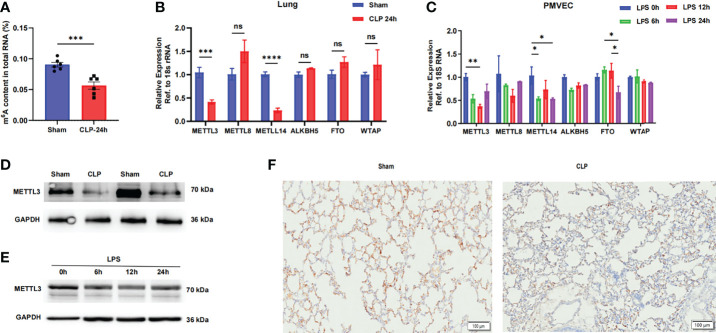
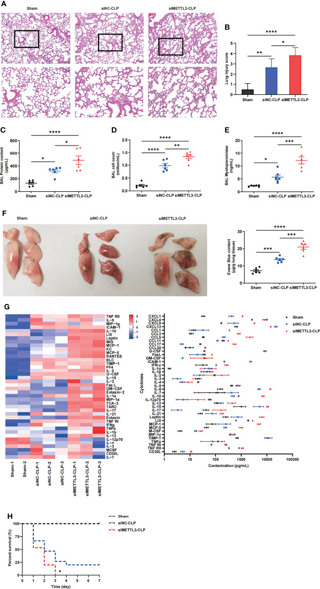
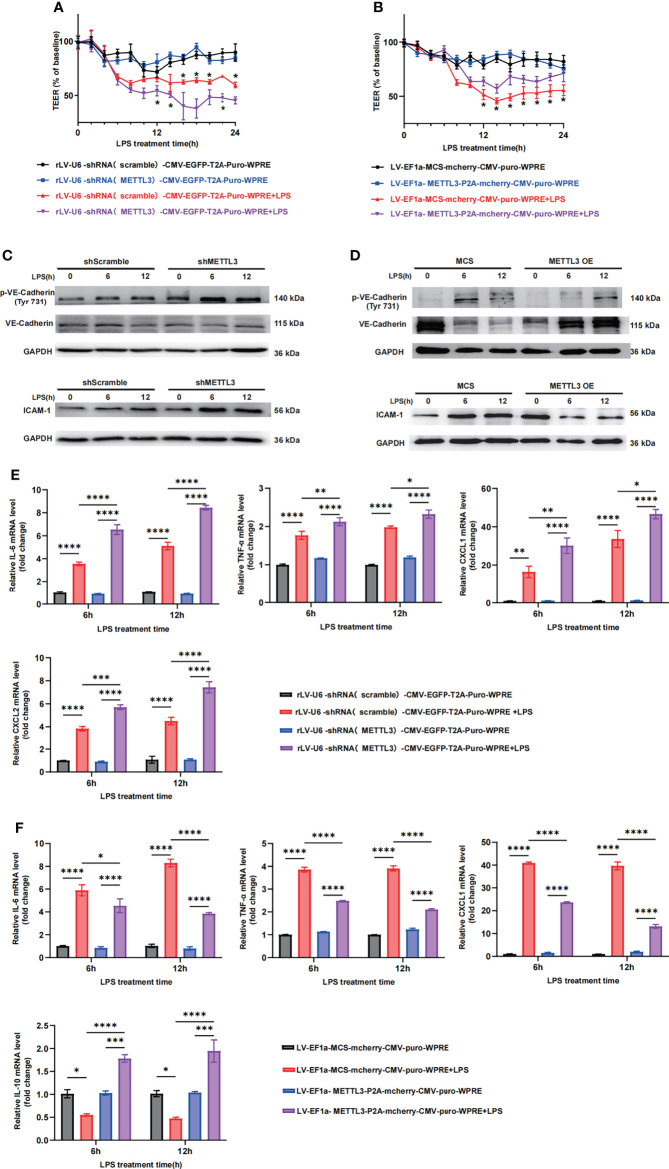
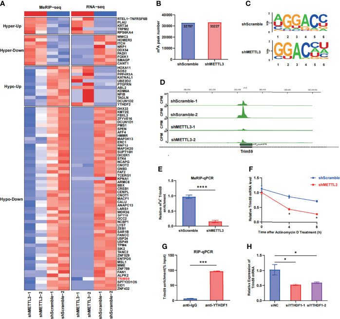
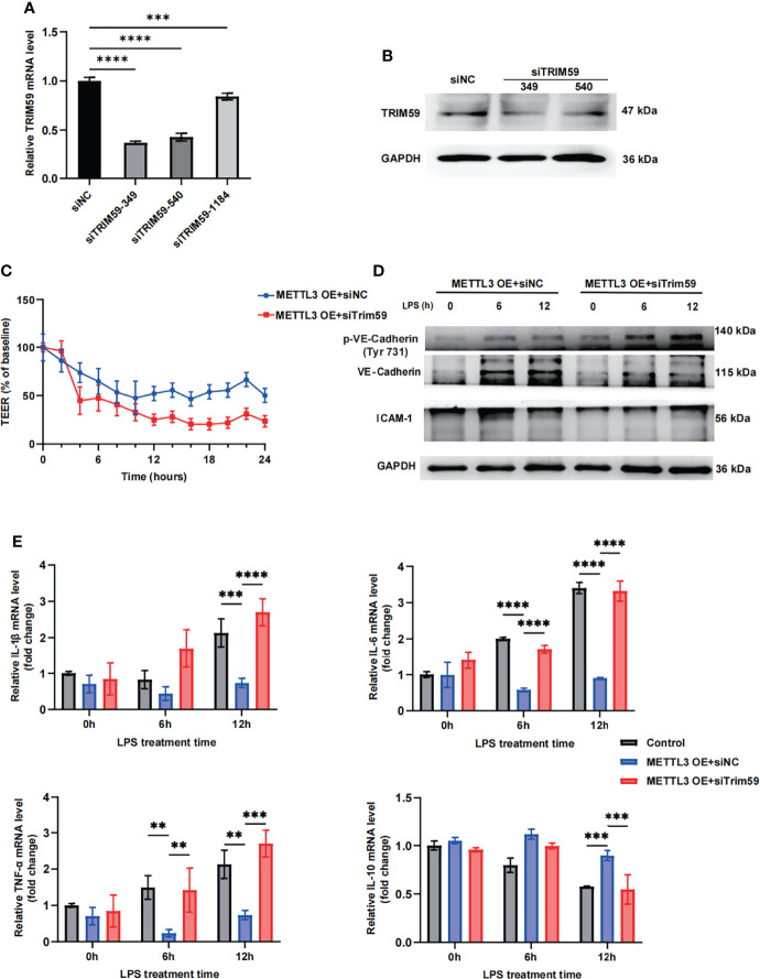
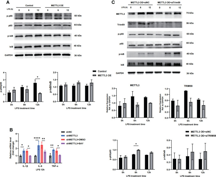
N6-methyladenosine (m6A) RNA modification is a fundamental determinant of mRNA metabolism in eukaryotic cells and is involved in numerous physiological and pathological processes. However, the specific role of m6A modification in sepsis-induced acute respiratory distress syndrome(ARDS) remains unknown. Here, we show that the levels of m6A RNA were significantly decreased in septic lungs and that METTL3 was the main regulator involved in the absence of m6A RNA modification. Pulmonary endothelial barrier damage is a critical process in the pathogenesis of acute lung injury during sepsis. METTL3 regulated endothelial barrier dysfunction and inflammatory responses in sepsis-induced ARDS in vivo and in vitro. Furthermore, we identified tripartite motif-containing (Trim)59 as a key m6A effector and Trim59 deficiency exacerbated lung injury. Mechanistically, METTL3 inhibited endothelial injury in sepsis-induced ARDS through Trim59-associated NF-κB inactivation. Our findings revealed novel insights into epitranscriptional mechanisms in sepsis-induced ARDS via m6A modifications, which has important application value in the diagnosis, prognosis, and molecular-targeted therapy of sepsis-associated lung injury.

Keywords: METTL3; acute lung injury; endothelial barrier; epigenetic regulation; m6A; sepsis.


Conflict of interest statement

The authors declare that the research was conducted in the absence of any commercial or financial relationships that could be construed as a potential conflict of interest.

Figures

Figure 1
Figure 1
The METTL3-m6A pathway is downregulated in sepsis-induced ARDS. (A) ELISA was used to measure mRNA m6A levels in sham and CLP-24 h lungs. (B, C) RT-qPCR analysis showing the mRNA expression of m6A-related genes in mouse lungs and PMVECs. 18S rRNA was used as the internal control. (D, E) Immunoblot showing METTL3 protein expression in mouse lungs and PMVECs. GAPDH was used as the loading control. (F) Representative images of METTL3 immunostaining in sham lungs and CLP lungs. Scale bars, 100 mm. All data are expressed as the mean ± SD of three independent experiments (n=6). *p < 0.05; **p<0.01; ***p < 0.001; ****p < 0.0001; ns, no significance.
Figure 2
Figure 2
METTL3 deletion exacerbates lung endothelial injury in CLP models. (A) Hematoxylin and eosin (H&E)-stained lung sections from sham, CLP-NC siRNA and CLP-METTL3 siRNA mice. Scale bars, 100 μm. (B) Pathological lung injury scores of sham, CLP-NC siRNA and CLP-METTL3 siRNA mice. (C–E) Protein content (C), cell counts (D) and MPO activity (E) in the BALF of CLP-METTL3 siRNA mice compared with sham and CLP-NC siRNA mice. (F) Whole lung images and Evans blue levels in sham, CLP-NC siRNA and CLP-METTL3 siRNA mice. (G) Quantibody Mouse Inflammation Array analysis of circulating blood from sham, CLP-NC siRNA and CLP-METTL3 siRNA mice. (H) Survival analysis of CLP-METTL3 siRNA mice compared with sham and CLP-NC siRNA mice. All data are expressed as the mean ± SD of three independent experiments (n=6 for A-G and n=15 for H). *p < 0.05; **p < 0.01; ***p < 0.001; ****p < 0.0001.
Figure 3
Figure 3
METTL3 regulates vascular endothelial barrier function in vitro. (A, B) TEER was used to measure endothelial permeability in HULEC-5a cells transfected with scramble or METTL3 shRNA (A) and in HULEC-5a cells with or without METTL3 overexpression (B). (C, D) Immunoblot showing the protein expression of VE-Cadherin and ICAM-1 in HULEC-5a cells transfected with scramble or METTL3 shRNA (C) and in HULEC-5a cells with or without METTL3 overexpression (D). (E, F) RT-qPCR analysis showing inflammatory cytokine mRNA expression in HULEC-5a cells transfected with scramble or METTL3 shRNA (E) and in HULEC-5a cells with or without METTL3 overexpression (F). All data are expressed as the mean ± SD of three independent experiments. *p < 0.05; **p < 0.01; ***p < 0.001; ****p < 0.0001.
Figure 4
Figure 4
METTL3-mediated m6A modification of Trim59 mRNA maintains its YTHDF1-dependent stability (A) Heatmap showing the clusters with significant changes in both the RNA expression level (RNA-seq) and m6A level (MeRIP-seq) in scramble and METTL3 shRNA-treated HULEC-5a cells. (B) Number of m6A peaks in scramble and METTL3 shRNA-treated HULEC-5a cells. (C) Global profiling of m6A in HULEC-5a cells and the sequence motif identified from the top 1000 m6A peaks. (D) MeRIP-seq plotted the m6A abundances on Trim59 mRNA transcripts in HULEC-5a cells. (E) MeRIP-qPCR examined METTL3-mediated Trim59 mRNA m6A modifications. (F) RT-qPCR analysis showing the levels of Trim59 expression in METTL3-downregulated HULEC-5a cells treated with actinomycin D (2 μg/mL) at the indicated time points. (G) RNA immunoprecipitation (RIP)-qPCR assay showing the enrichment of YTHDF1 binding to Trim59 m6A modification sites. (H) RT-qPCR analysis showing the levels of Trim59 expression in YTHDF1-knockdown HULEC-5a cells. All data are expressed as the mean ± SD of three independent experiments. *p < 0.05; ***p < 0.001; ****p < 0.0001.
Figure 5
Figure 5
METTL3 regulates endothelial function by targeting Trim59. (A) RT-qPCR analysis showing the levels of Trim59 expression after siRNA transfection. (B) Immunoblot showing Trim59 protein expression after siRNA transfection. (C) TEER was used to measure endothelial permeability in METTL3-overexpressing HULEC-5a cells with or without Trim59 siRNA. (D) Immunoblot showing the protein expression of VE-Cadherin and ICAM-1 in METTL3-overexpressing HULEC-5a cells with or without Trim59 siRNA. (E) RT-qPCR analysis showing inflammatory cytokine mRNA expression in METTL3-overexpressing HULEC-5a cells with or without Trim59 siRNA. All data are expressed as the mean ± SD of three independent experiment. **p < 0.01; ***p < 0.001; ****p < 0.0001.
Figure 6
Figure 6
METTL3 inhibits endothelial injury in sepsis-induced ARDS through Trim59-associated NF-κB inactivation. (A, C) Immunoblot showing p65 and IκB phosphorylation in HULEC-5a cells treated with or without METTL3 overexpression (A) and METTL3-overexpressing HULEC-5a cells with or without Trim59 siRNA (C). (B) RT-qPCR analysis showing inflammatory cytokine and endothelial functional marker mRNA expression in METTL3-overexpressing HULEC-5a cells treated with or without the NF-κB inhibitor BAY. All data are expressed as the mean ± SD of three independent experiments. *p < 0.05; **p < 0.01; ****p < 0.0001.

Similar articles

Cited by

References

    1. Singer M, Deutschman CS, Seymour CW, Shankar-Hari M, Annane D, Bauer M, et al. . The Third International Consensus Definitions for Sepsis and Septic Shock (Sepsis-3). Jama-J Am Med Assoc (2016) 315(8):801–10. doi: 10.1001/jama.2016.0287 - DOI - PMC - PubMed
    1. Matthay MA, Zemans RL, Zimmerman GA, Arabi YM, Beitler JR, Mercat A, et al. . Acute Respiratory Distress Syndrome. Nat Rev Dis Primers (2019) 5 5(1):18. doi: 10.1038/s41572-019-0069-0 - DOI - PMC - PubMed
    1. Spinelli E, Mauri T, Beitler JR, Pesenti A, Brodie D. Respiratory Drive in the Acute Respiratory Distress Syndrome: Pathophysiology, Monitoring, and Therapeutic Interventions. Intensive Care Med (2020) 46(4):606–18. doi: 10.1007/s00134-020-05942-6 - DOI - PMC - PubMed
    1. Huppert LA, Matthay MA, Ware LB. Pathogenesis of Acute Respiratory Distress Syndrome. Semin Respir Crit Care Med (2019) 40(1):31–9. doi: 10.1055/s-0039-1683996 - DOI - PMC - PubMed
    1. Joffre J, Hellman J, Ince C, Ait-Oufella H. Endothelial Responses in Sepsis. Am J Respir Crit Care Med (2020) 202(3):361–70. doi: 10.1164/rccm.201910-1911TR - DOI - PubMed

Publication types

MeSH terms