Review
. 2022 May 27:13:857954.
doi: 10.3389/fimmu.2022.857954. eCollection 2022.

Conventional Type 1 Dendritic Cells in Intestinal Immune Homeostasis

Affiliations
Review

Conventional Type 1 Dendritic Cells in Intestinal Immune Homeostasis

Izumi Sasaki et al. Front Immunol. .

Abstract

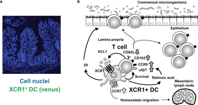
Dendritic cells (DC) play critical roles in linking innate and adaptive immunity. DC are heterogenous and there are subsets with various distinct functions. One DC subset, conventional type 1 DC (cDC1), can be defined by expression of CD8α/CD103 in mice and CD141 in humans, or by expression of a chemokine receptor, XCR1, which is a conserved marker in both mice and human. cDC1 are characterized by high ability to ingest dying cells and to cross-present antigens for generating cytotoxic CD8 T cell responses. Through these activities, cDC1 play crucial roles in immune responses against infectious pathogens or tumors. Meanwhile, cDC1 involvement in homeostatic situations is not fully understood. Analyses by using mutant mice, in which cDC1 are ablated in vivo, revealed that cDC1 are critical for maintaining intestinal immune homeostasis. Here, we review the homeostatic roles of cDC1, focusing upon intestinal immunity.

Keywords: T cell; XCR1; dendritic cell; gene targeting; intestine; subset.


Conflict of interest statement

The authors declare that the research was conducted in the absence of any commercial or financial relationships that could be construed as a potential conflict of interest.

Figures

Figure 1
Figure 1
Crosstalk between XCR1+ DC and intestinal T cells. (A) Immunofluorescence imaging of intestinal sections from XCR1-Venus (Xcr1+/venus ) mice. XCR1+ DC can be detected by Venus expression. Cell nuclei were stained with Diamidino-2-phenylindole. (B) Once activated, intestinal T cells produce XCL1, which attracts XCR1+ DC. XCR1+ DC support survival, upregulation of CD103, CCR9, α4β7, and XCL1 expression, downregulation of CD62L expression and generation of CD4+CD8αα+ IEL, thereby leading to maintenance of intraepithelial and LP T cell populations. T cell-derived XCL1 then keep CCR7 expression of XCR1+ DC to enable migration of XCR1+ DC from the LP to the mesenteric lymph nodes. High expression of XCR1 and β8 integrin and high activity of aldehyde dehydrogenase, which can convert retinal to retinoic acid, contribute to XCR1+DC-dependent mechanisms.

References

    1. Mowat AM, Agace WW. Regional Specialization Within the Intestinal Immune System. Nat Rev Immunol (2014) 14:667–85. doi: 10.1038/nri3738 - DOI - PubMed
    1. Steinman RM. Decisions About Dendritic Cells: Past, Present, and Future. Annu Rev Immunol (2012) 30:1–22. doi: 10.1146/annurev-immunol-100311-102839 - DOI - PubMed
    1. Liu K, Nussenzweig MC. Origin and Development of Dendritic Cells. Immunol Rev (2010) 234:45–54. doi: 10.1111/j.0105-2896.2009.00879.x - DOI - PubMed
    1. Merad M, Sathe P, Helft J, Miller J, Mortha A. The Dendritic Cell Lineage: Ontogeny and Function of Dendritic Cells and Their Subsets in the Steady State and the Inflamed Setting. Annu Rev Immunol (2013) 31:563–604. doi: 10.1146/annurev-immunol-020711-074950 - DOI - PMC - PubMed
    1. Audsley KM, McDonnell AM, Waithman J. Cross-Presenting XCR1(+) Dendritic Cells as Targets for Cancer Immunotherapy. Cells (2020) 9:565. doi: 10.3390/cells9030565 - DOI - PMC - PubMed

Publication types

Substances