Review
. 2022 May 25:13:914033.
doi: 10.3389/fimmu.2022.914033. eCollection 2022.

Myocardial-Treg Crosstalk: How to Tame a Wolf

Affiliations
Review

Myocardial-Treg Crosstalk: How to Tame a Wolf

Emil Weiß et al. Front Immunol. .

Abstract

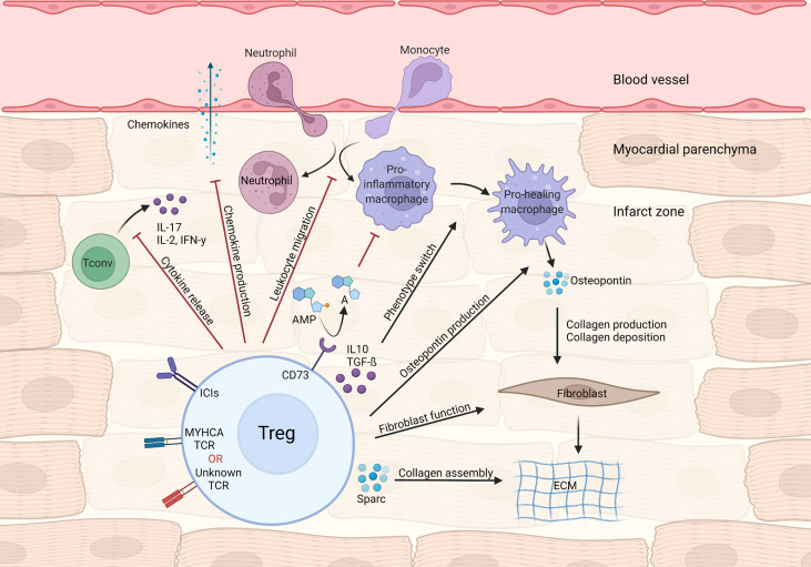
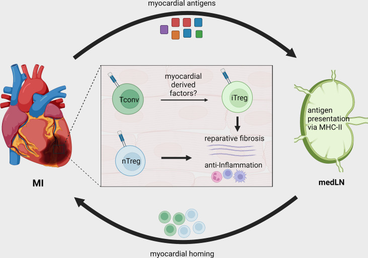
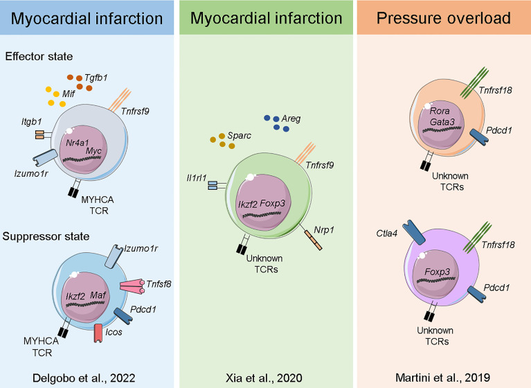
The immune system plays a vital role in maintaining tissue integrity and organismal homeostasis. The sudden stress caused by myocardial infarction (MI) poses a significant challenge for the immune system: it must quickly substitute dead myocardial with fibrotic tissue while controlling overt inflammatory responses. In this review, we will discuss the central role of myocardial regulatory T-cells (Tregs) in orchestrating tissue repair processes and controlling local inflammation in the context of MI. We herein compile recent advances enabled by the use of transgenic mouse models with defined cardiac antigen specificity, explore whole-heart imaging techniques, outline clinical studies and summarize deep-phenotyping conducted by independent labs using single-cell transcriptomics and T-cell repertoire analysis. Furthermore, we point to multiple mechanisms and cell types targeted by Tregs in the infarcted heart, ranging from pro-fibrotic responses in mesenchymal cells to local immune modulation in myeloid and lymphoid lineages. We also discuss how both cardiac-specific and polyclonal Tregs participate in MI repair. In addition, we consider intriguing novel evidence on how the myocardial milieu takes control of potentially auto-aggressive local immune reactions by shaping myosin-specific T-cell development towards a regulatory phenotype. Finally, we examine the potential use of Treg manipulating drugs in the clinic after MI.

Keywords: Foxp3; T-cells; Tregs (regulatory T cells); fibrosis; heart; myocardial infarction.


Conflict of interest statement

The authors declare that the research was conducted in the absence of any commercial or financial relationships that could be construed as a potential conflict of interest.

Figures

Figure 1
Figure 1
Treg-mediated effects in myocardial repair and inflammation after infarction. Tregs control local immune responses through a multitude of mechanisms, including inhibiting both canonical TH1/TH17 cytokine production and leukocyte migration. Treg cytokines (e.g. IL-10 and TGF-β) and purinergic metabolism products may modulate macrophage polarization towards a pro-healing/reparative phenotype. In addition, Tregs contribute to fibroblast activation and steady collagen deposition in the infarcted area.
Figure 2
Figure 2
Treg recruitment and in situ conversion during MI. After MI, cardiac autoantigens, including MYHCA, are drained to local MedLN and presented to CD4+ T-cells via the MHC-II molecule. Naturally existing thymus derived Tregs (nTreg) and conventional T-cells are activated in the MedLN after MI and migrate to the infarcted tissue. The myocardial immune crosstalk induces myosin-specific T-cells to acquire an induced regulatory phenotype (iTreg). Both induced and naturally occurring Tregs contribute to tissue repair by modulating local inflammatory response and fostering tissue fibrosis.
Figure 3
Figure 3
Transcriptome of myocardial Tregs in the injured heart. Single-cell sequencing of heart and MedLN T-cells after MI (40) Delgobo et al., 2022 revealed that TCR-M cells have an induced Treg signature characterized by effector-state Tregs expressing high levels of Tgfb1 and suppressor cells expressing several immune checkpoint receptors (e.g. Pdcd1, Icos, Tigit). Single-cell and bulk RNA sequencing of cardiac T-cells after MI (44) Xia et al., 2020 showed Treg clonal expansion, a Treg thymus-derived signature and the production of pro-healing transcripts such as Areg and Sparc. Single-cell sequencing of myocardial leukocytes in a pressure overload model (48) Martini et al., 2019 demonstrated that two main Treg populations expanded one week post injury. Both bona fide Tregs and those with TH17/non-lymphoid gene signatures were identified, and both expressed high levels of Pdcd1 immune checkpoint receptor.

References

    1. Swirski FK, Nahrendorf M. Cardioimmunology: The Immune System in Cardiac Homeostasis and Disease. Nat Rev Immunol (2018) 18(12):733–44. doi: 10.1038/s41577-018-0065-8 - DOI - PubMed
    1. Hulsmans M, Clauss S, Xiao L, Aguirre AD, King KR, Hanley A, et al. . Macrophages Facilitate Electrical Conduction in the Heart. Cell (2017) 169(3):510–22 e20. doi: 10.1016/j.cell.2017.03.050 - DOI - PMC - PubMed
    1. Hofmann U, Frantz S. Role of T-cells in Myocardial Infarction. Eur Heart J (2016) 37(11):873–9. doi: 10.1093/eurheartj/ehv639 - DOI - PubMed
    1. Nevers T, Salvador AM, Grodecki-Pena A, Knapp A, Velazquez F, Aronovitz M, et al. . Left Ventricular T-Cell Recruitment Contributes to the Pathogenesis of Heart Failure. Circ Heart Fail (2015) 8(4):776–87. doi: 10.1161/CIRCHEARTFAILURE.115.002225 - DOI - PMC - PubMed
    1. Kallikourdis M, Martini E, Carullo P, Sardi C, Roselli G, Greco CM, et al. . T Cell Costimulation Blockade Blunts Pressure Overload-Induced Heart Failure. Nat Commun (2017) 8:14680. doi: 10.1038/ncomms14680 - DOI - PMC - PubMed

Publication types