

The Effect of Self-Myofascial Release on the Pennation Angle of the Vastus Medialis Oblique and the Vastus Lateralis in Athletic Male Individuals: An Ultrasound Investigation
- PMID: 35693863
- PMCID: PMC9159710
- DOI: 10.26603/001c.35591
The Effect of Self-Myofascial Release on the Pennation Angle of the Vastus Medialis Oblique and the Vastus Lateralis in Athletic Male Individuals: An Ultrasound Investigation
Abstract
Background: Maintenance of patellar stability requires a balance between the vastus medialis oblique (VMO) and the vastus lateralis (VL). The imbalance between these muscles is thought to be implicated in the etiology of patellofemoral pain (PFP). Where there is hypertrophy of the VL in PFP patients, self-myofascial release (SMR) may be utilized for its management. However, there is no current evidence regarding SMR and its effects on VMO and VL architecture. The aim of this study, therefore, was to use ultrasound to gain further understanding of the effects of a program of SMR on the fiber angles of the VMO and VL.
Hypothesis: There will be a significant decrease in the pennation angles of the VMO and VL after seven weeks of SMR using a foam roller.
Study design: Cohort Study.
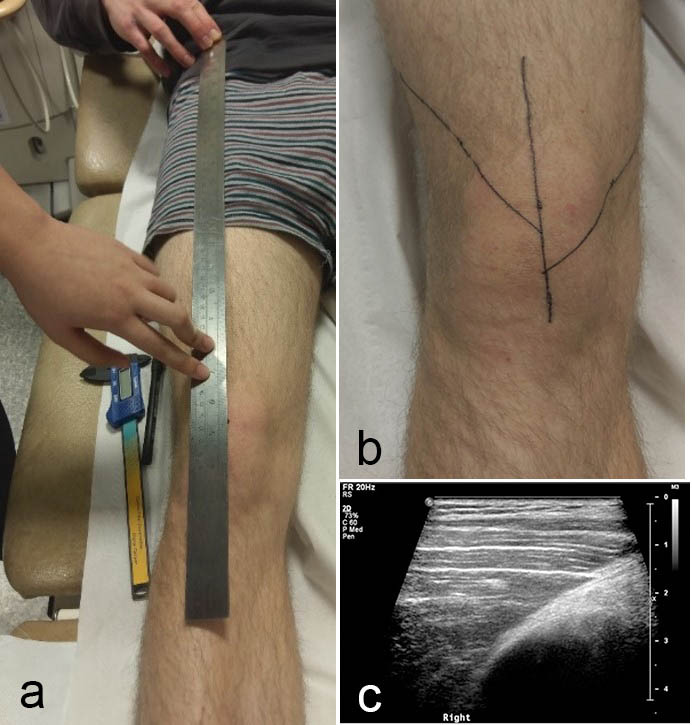
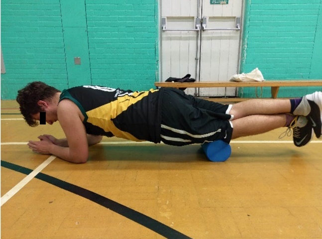
Methods: Twenty-five young, athletic, male participants were recruited to use a foam roller, along the full length of both anterior thighs, three times weekly, on three separate days, for seven weeks. Ultrasound was used to determine the initial and final VMO and VL pennation angles on both limbs. One eligible participant was chosen as an intra-rater control and did not partake in the SMR regimen.
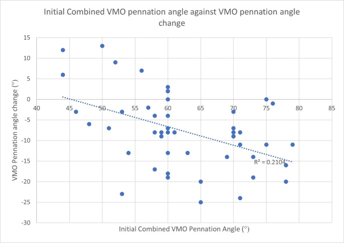
Results: There was a statistically significant (p < 0.001) decrease in the pennation angles of the VMO and VL after the SMR regime. Mean combined right and left VL angle change was -6.65° (-18% mean change) and the mean combined right and left VMO angle change was -7.65° (-11.5% mean change). A weak negative correlation was found between initial VMO fiber angle and the angle change (Rsquared = -0.21), as well as moderate negative correlation for the VL (Rsquared = -0.51).
Conclusion: A program of SMR on the anterior thighs of young, asymptomatic males resulted in changes to the fiber angles of both the VMO and VL. There was a significant decrease in pennation angle after seven weeks of SMR using a foam roller.
Keywords: Patellofemoral pain; physiotherapy; self-myofascial release; ultrasound; vastus lateralis; vastus medialis oblique.
Figures

References
-
- Vastus medialis: a reappraisal of VMO and VML. Skinner Emily J, Adds Philip J. 2012Journal of Physical Therapy Science. 24(6):475–479. doi: 10.1589/jpts.24.475. doi: 10.1589/jpts.24.475. - DOI
LinkOut - more resources
Full Text Sources
Medical