. 2022 Apr 20;13(19):5760-5766.
doi: 10.1039/d1sc07151c. eCollection 2022 May 18.

Discovery of a phase-separating small molecule that selectively sequesters tubulin in cells

Affiliations

Discovery of a phase-separating small molecule that selectively sequesters tubulin in cells

Genyir Ado et al. Chem Sci. .

Abstract

Phase-separated membraneless organelles or biomolecular condensates play diverse functions in cells, however recapturing their characteristics using small organic molecules has been a challenge. In the present study, cell-lysate-based screening of 843 self-assembling small molecules led to the discovery of a simple organic molecule, named huezole, that forms liquid droplets to selectively sequester tubulin. Remarkably, this small molecule enters cultured human cells and prevents cell mitosis by forming tubulin-concentrating condensates in cells. The present study demonstrates the feasibility of producing a synthetic condensate out of non-peptidic small molecules for exogenous control of cellular processes. The modular structure of huezole provides a framework for designing a class of organelle-emulating small molecules.


Conflict of interest statement

There are no conflicts to declare.

Figures
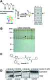
Fig. 1
Fig. 1. Discovery of huezole. (A) A schematic diagram of the screening procedure. (B) A representative silver-stained image of a screening gel of self-assembly library compounds. The protein band specific for huezole (1) is marked with an arrow. (C) Chemical structure of huezole. (D) Dose-dependent co-precipitation of tubulin with huezole. Immunoblots of supernatant and pellet fractions with indicated antibodies are shown.
Fig. 2
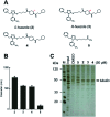
Fig. 2. Self-assembling properties of huezole analogues 2–4. (A) Chemical structures of huezole analogues 2–5. (B) Average hydrodynamic diameters of the compounds at 50 μM in PBS. (C) Silver-stained gel image showing the co-precipitation of tubulin with analogues 2–5 in cell lysates.
Fig. 3
Fig. 3. Liquid-like phase separation of R-huezole (3). (A) Bright field time-lapse images of coalescing puncta of R-huezole (3). Scale bars, 5 μm. (B) FRAP assays of Nile-Red-doped R-huezole particles (90 nM Nile Red and 50 μM R-huezole). Recovery of the normalized fluorescence intensities indicated t1/2 values in the absence (9.88 s) and presence (20.3 s) of tubulin (200 nM). The blue lines indicate SEM, and the red lines show the results of exponential fitting with the average intensities of fluorescent particles (N = 30 for Nile Red + R-huezole and N = 94 for Nile Red + R-huezole + tubulin, respectively).
Fig. 4
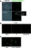
Fig. 4. Microscopic observation of the effects of R-huezole (3) on tubulin in vitro. (A) In vitro tubulin sequestration with R-huezole (3). Labeled tubulin (200 nM) was incubated with DMSO (1%) or R-huezole (50 μM) for 30 min before imaging. Scale bars, 10 μm. (B) Effects of R-huezole (3) on Taxol-induced tubulin polymerization in vitro. Labeled tubulin was dissolved in G-PEM buffer including Taxol (1 μM) followed by incubation with 50 μM R-huezole (3) for 30 min at 37 °C prior to imaging. Scale bars, 20 μm. (C) R-huezole-tubulin condensates under a highly polymerization-inducing condition. Polymerization of labeled tubulin (900 nM) was induced overnight by Taxol (50 μM) in the presence or absence of 50 μM R-huezole (3) in PBS. Scale bar, 5 μm.
Fig. 5
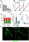
Fig. 5. Effects of R-huezole (3) in cells. (A) HeLa cells were treated with DMSO (0.5%), 5 (50 μM), or R-huezole (10 and 20 μM). Cell viability was monitored by WST-8 assay after 48 h. (B) R-huezole (3) delays the proliferation of HeLa cells. Passage-based cell proliferation was monitored at 4, 8, or 12 days after treatment with DMSO or R-huezole (5 μM). (C) R-huezole (3) treatment leads to G2/M cell-cycle arrest. HEK293 cells were transfected with Fucci fluorescent probes mCherry-hCdt1 (30/120) (red) and AmCyan-hGeminin (green) followed by treatment with DMSO or R-huezole (3). An increase in AmCyan-hGeminin (green) fluorescence indicates an increase in G2/M cell population. Nocodazole (1 μM) was used as a positive control. (D) Effects of R-huezole (3) on mitotic index (% of cells with condensed chromatin). HEK293 cells were stained for nucleus with Hoechst 33342 after 48 h treatment with R-huezole. Data represent average values mean ± S.D (n = 3). The p values (*p < 0.05 and ****p < 0.0001) were determined using one-way ANOVA followed by Dunnett's multiple comparisons test with a 95% confidence interval. (E) Cellular tubulin sequestration with R-huezole (3). HeLa cells were transduced with CellLight™ tubulin-GFP followed by treatment with DMSO or R-huezole (50 μM) for 3 h prior to imaging. HeLa cells were analysed for puncta and 5 cells from at least 7 microscopic fields were quantified with similar results from three independent experiments. Data represent average values mean ± S.D (n = 3). The p value (*p < 0.05) was determined by an unpaired two-tailed Student's t-test. (F) Representative confocal images of the 3-induced cellular tubulin sequestration. DMSO-treated cells (left) show uniform tubulin-GFP expression; R-huezole treated cells (right) show intracellular puncta indicating sequestration with R-huezole.

References

    1. Shin Y. Brangwynne C. P. Science. 2017;357:eaaf4382. doi: 10.1126/science.aaf4382. - DOI - PubMed
    1. Banani S. F. Lee H. O. Hyman A. A. Rosen M. K. Nat. Rev. Mol. Cell Biol. 2017;18:285–298. doi: 10.1038/nrm.2017.7. - DOI - PMC - PubMed
    1. O'Flynn B. G. Mittag T. Curr. Opin. Cell Biol. 2021;69:70–79. doi: 10.1016/j.ceb.2020.12.012. - DOI - PMC - PubMed
    1. Hnisz D. Shrinivas K. Young R. A. Chakraborty A. K. Sharp P. A. Cell. 2017;169:13–23. doi: 10.1016/j.cell.2017.02.007. - DOI - PMC - PubMed
    1. Ditlev J. A. Case L. B. Rosen M. K. J. Mol. Biol. 2018;430:4666–4684. doi: 10.1016/j.jmb.2018.08.003. - DOI - PMC - PubMed