. 2022 May 25:9:817866.
doi: 10.3389/fcvm.2022.817866. eCollection 2022.

Isolated Coarctation of the Aorta: Current Concepts and Perspectives

Affiliations

Isolated Coarctation of the Aorta: Current Concepts and Perspectives

Ami B Bhatt et al. Front Cardiovasc Med. .

Abstract

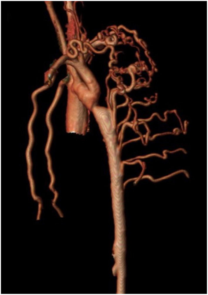
Current management of isolated CoA, localized narrowing of the aortic arch in the absence of other congenital heart disease, is a success story with improved prenatal diagnosis, high survival and improved understanding of long-term complication. Isolated CoA has heterogenous presentations, complex etiologic mechanisms, and progressive pathophysiologic changes that influence outcome. End-to-end or extended end-to-end anastomosis are the favored surgical approaches for isolated CoA in infants and transcatheter intervention is favored for children and adults. Primary stent placement is the procedure of choice in larger children and adults. Most adults with treated isolated CoA thrive, have normal daily activities, and undergo successful childbirth. Fetal echocardiography is the cornerstone of prenatal counseling and genetic testing is recommended. Advanced 3D imaging identifies aortic complications and myocardial dysfunction and guides individualized therapies including re-intervention. Adult CHD program enrollment is recommended. Longer follow-up data are needed to determine the frequency and severity of aneurysm formation, myocardial dysfunction, and whether childhood lifestyle modifications reduce late-onset complications.

Keywords: adult congenital heart disease; catheter intervention; coarctation of the aorta; congenital heart disease; heart surgery.


Conflict of interest statement

The authors declare that the research was conducted in the absence of any commercial or financial relationships that could be construed as a potential conflict of interest.

Figures

Figure 1
Figure 1
(A,B) Echocardiographic images of Isolated CoA. (A) Shows B-Mode and color Doppler images of discrete isthmus CoA with obvious size discrepancy between the isthmus and PDA diameters. (B) Shows an abnormal abdominal aorta Doppler profile with a low velocity signal, blunted upstroke with delayed systolic peaking, a slurred downstroke and continuous diastolic flow in a patient with isolated CoA.
Figure 2
Figure 2
Cardiac magnetic resonance imaging (MRI) of isolated CoA. Cardiac MRI showing a discrete and isolated CoA. This modality offers high resolution imaging of the entire aortic arch, helping localize the extent and significance of the coarctation.
Figure 3
Figure 3
Cardiac magnetic resonance angiography (MRA) of isolated CoA with 4D flow. Advanced phase contrast magnetic resonance angiograms (PC-MRA) allows for both 3-dimensional anatomic evaluation of arch anatomy as well as 4-dimensional blood flow visualization using time resolved streamlines. Image courtesy of Joshua Robinson and Michael Markl (Dept. of Radiology, Northwestern University).
Figure 4
Figure 4
Cardiac magnetic resonance angiography (MRA) demonstrating isolated CoA with prominent collateralization. A patient with isolated CoA and prominent collateral vessel formation.
Figure 5
Figure 5
Cardiac computed tomography (CT) imaging of isolated CoA following stent implantation. Cardiac CT also allows for high resolution imaging of the entire aortic arch and enables visualization of possible in-stent stenosis.
Figure 6
Figure 6
Cardiac magnetic resonance imaging after isolated CoA repair with aneurysm formation. 3D reconstruction of a patient's aorta with a Gothic arch and aneurysm formation at the site of prior isolated CoA repair.
Figure 7
Figure 7
Central illustration: continuum of care for isolated coarctation of the aorta. The continuum of care for isolated coarctation of the aorta includes clinical diagnostics (A); neurodevelopmental assessment (B); surgical (C) or percutaneous intervention (D); longitudinal care during adulthood (E); and pregnancy management including genetic counseling (F). Individualized management strategies are determined by age at presentation, anatomic features of CoA, and associated conditions including other CHD and genetic syndromes.

References

    1. Raissadati A, Nieminen H, Haukka J, Sairanen H, Jokinen E. Late causes of death after pediatric cardiac surgery: a 60-year population-based study. J Am Coll Cardiol. (2016) 68:487–98. 10.1016/j.jacc.2016.05.038 - DOI - PubMed
    1. Kappetein AP. Gittenberger-de Groot AC, Zwinderman AH, Rohmer J, Poelmann RE, Huysmans HA. The neural crest as a possible pathogenetic factor in coarctation of the aorta and bicuspid aortic valve. J Thorac Cardiovasc Surg. (1991) 102:830–6. 10.1016/S0022-5223(19)33931-5 - DOI - PubMed
    1. Gittenberger-de Groot AC, Azhar M, Molin DG. Transforming growth factor beta-SMAD2 signaling and aortic arch development. Trends Cardiovasc Med. (2006) 16:1–6. 10.1016/j.tcm.2005.09.006 - DOI - PubMed
    1. Ho SY, Anderson RH. Coarctation, tubular hypoplasia, and the ductus arteriosus. Histological study of 35 specimens. Br Heart J. (1979) 41:268–74. 10.1136/hrt.41.3.268 - DOI - PMC - PubMed
    1. Pfaltzgraff ER, Shelton EL, Galindo CL, Nelms BL, Hooper CW, Poole SD, et al. . Embryonic domains of the aorta derived from diverse origins exhibit distinct properties that converge into a common phenotype in the adult. J Mol Cell Cardiol. (2014) 69:88–96. 10.1016/j.yjmcc.2014.01.016 - DOI - PMC - PubMed

Publication types

LinkOut - more resources