. 2022 Jun 21;119(25):e2202932119.
doi: 10.1073/pnas.2202932119. Epub 2022 Jun 13.

Essential functions of mosquito ecdysone importers in development and reproduction

Affiliations

Essential functions of mosquito ecdysone importers in development and reproduction

Lewis V Hun et al. Proc Natl Acad Sci U S A. .

Abstract

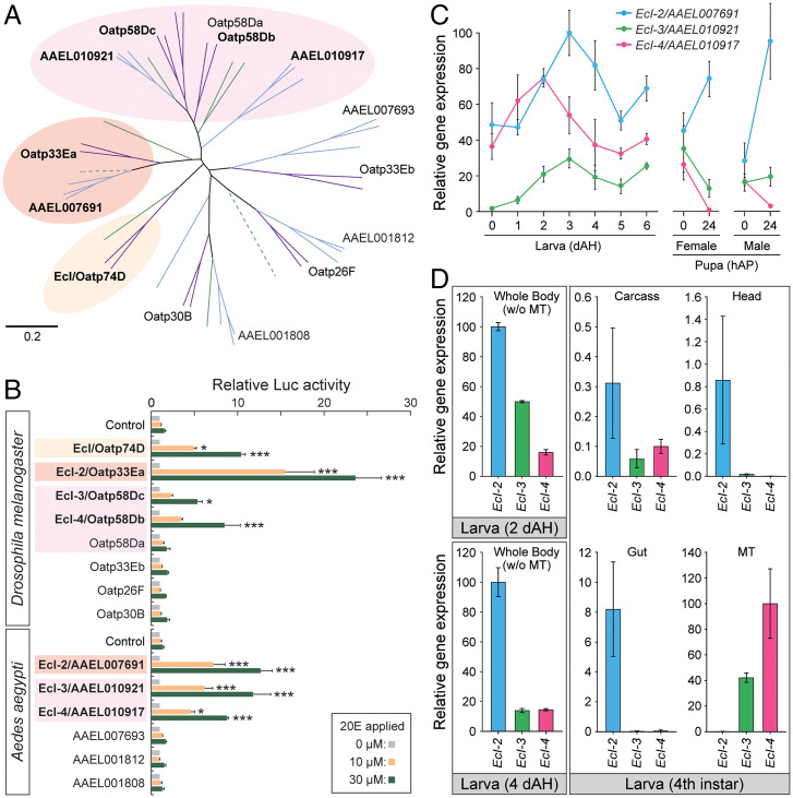
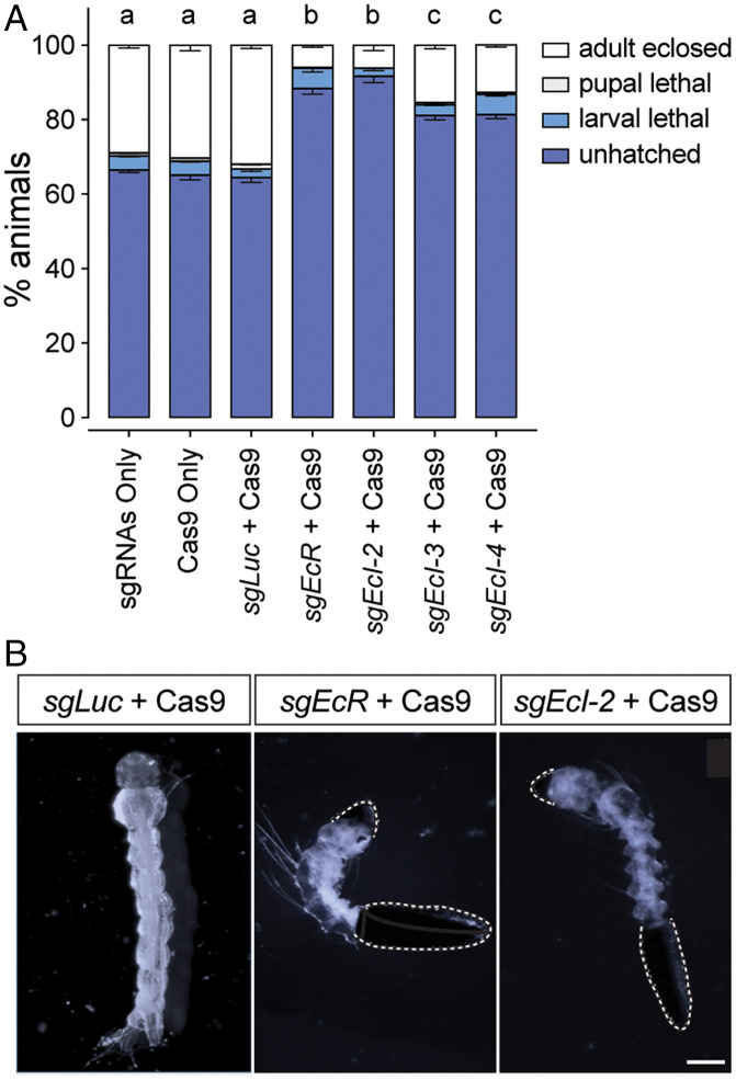
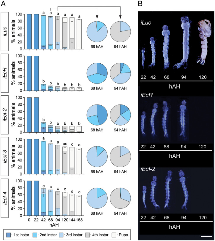
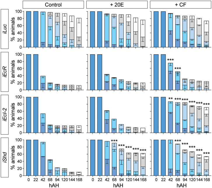
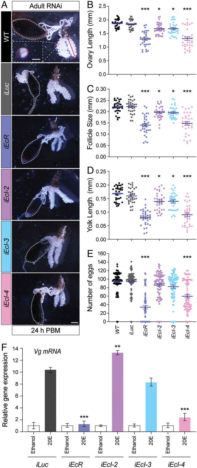
The primary insect steroid hormone ecdysone requires a membrane transporter to enter its target cells. Although an organic anion-transporting polypeptide (OATP) named Ecdysone Importer (EcI) serves this role in the fruit fly Drosophila melanogaster and most likely in other arthropod species, this highly conserved transporter is apparently missing in mosquitoes. Here we report three additional OATPs that facilitate cellular incorporation of ecdysone in Drosophila and the yellow fever mosquito Aedes aegypti. These additional ecdysone importers (EcI-2, -3, and -4) are dispensable for development and reproduction in Drosophila, consistent with the predominant role of EcI. In contrast, in Aedes, EcI-2 is indispensable for ecdysone-mediated development, whereas EcI-4 is critical for vitellogenesis induced by ecdysone in adult females. Altogether, our results indicate unique and essential functions of these additional ecdysone importers in mosquito development and reproduction, making them attractive molecular targets for species- and stage-specific control of ecdysone signaling in mosquitoes.

Keywords: Aedes aegypti; Drosophila melanogaster; ecdysone; organic anion-transporting polypeptide (OATP); vitellogenesis.


Conflict of interest statement

The authors declare no competing interest.

Figures

Fig. 1.
Fig. 1.
Identification of additional ecdysone importers in Drosophila and Aedes. (A) Neighbor-joining unrooted phylogenetic tree of full-length OATP proteins from representative dipteran insect species. Purple, flies (D. melanogaster and M. domestica); green, sand flies (P. papatasi); blue, mosquitoes (Ae. aegypti, A. gambiae, and Culex quinquefasciatus). Dotted lines indicate pseudogenes. Drosophila and Aedes OATPs are labeled. Protein names and GenBank accession numbers are listed in SI Appendix, Table S1. Scale bar indicates an evolutionary distance of 0.2 amino acid substitutions per position. (B) Luciferase (Luc) reporter activities in response to 10 μM or 30 μM 20E in HEK293 cells expressing Drosophila or Aedes OATPs. The cells were transfected with modified EcR (VgEcR) and RXR, along with each OATP-containing vector and luciferase reporter plasmids. Values are relative to the basal level (0 M 20E). All values are the means ± SEM (n = 2 to 4). *P < 0.05, ***P < 0.001 from one-way ANOVA followed by Dunnett’s multiple comparison test as compared to the response of the control cells to the same concentration of 20E. (C) Relative expression levels of ecdysone importer genes in the whole body during Aedes development, as assessed by qRT-PCR. Values are shown as percentages relative to the highest expression level of EcI-2. dAH, days after hatching, hAP, hours after pupation. All values are the means ± SEM (n = 3). (D) Tissue-specific expression of ecdysone importer genes during Aedes development, as assessed by qRT-PCR. Whole body samples without Malpighian tubules from early developmental stages (2 and 4 d after hatching) or individual tissues of fourth instar larvae were collected. Values are shown as percentages relative to the expression levels of EcI-2 in early developmental stages, or EcI-4 in the Malpighian tubule in the fourth instar. MT, Malpighian tubule. All values are the means ± SEM (n = 3).
Fig. 2.
Fig. 2.
Aedes ecdysone importers are required for embryogenesis. (A) Lethal stages of Aedes embryos injected with Cas9 protein and/or sgRNAs against control (sgLuc), EcR (sgEcR), or ecdysone importer genes (sgEcI-2, sgEcI-3, and sgEcI-4). Total 450 eggs were injected in three independent batches for each treatment, and their lethal stages were monitored thereafter. All values are the means ± SEM. Same letters on Top of the bars indicate statistically insignificant differences in embryonic lethality based on one-way ANOVA with Tukey's honestly significant difference (HSD) test. (B) Hatching defects observed in EcR and EcI-2 mutants. As compared to control (sgLuc + Cas9) that hatched normally, EcR or EcI-2 mutagenesis caused hatching defects in some larvae, where the eggshell remained attached (dotted white lines). (Scale bar, 0.5 mm.)
Fig. 3.
Fig. 3.
Aedes EcI-2 is required for larval developmental transitions. (A) Developmental progression and survival rate (%) of Luc RNAi (iLuc; control), EcR RNAi (iEcR), and EcI-2, -3, and -4 RNAi (iEcI-2, iEcI-3, and iEcI-4) animals. Color bars indicate developmental stages determined by stage-specific morphologic features such as the head capsule size and siphon length (42, 43). All values are the means ± SEM from seven independent experiments with 20 individuals in each replicate. hAH, hours after hatching. Between 42 and 168 hAH, same letters on Top of the bars indicate statistically insignificant differences in survival rate based on one-way ANOVA followed by Bonferroni’s multiple comparison test among RNAi animals at the same time point. Developmental stages of surviving larvae at 68 and 94 hAH are shown on the Right as pie charts. (B) Developmental progression of animals treated with dsRNA targeting Luc (iLuc; control), EcR (iEcR), or EcI-2 (iEcI-2). Representative images of animals at various time points were combined into single panels. (Scale bar, 1 mm.)
Fig. 4.
Fig. 4.
A nonsteroidal ecdysone agonist can rescue developmental arrest caused by EcI-2 knockdown in Aedes. Developmental progression and survival rate (%) of Luc RNAi (iLuc; control), EcR RNAi (iEcR), EcI-2 RNAi (iEcI-2), and shade RNAi (iShd) animals treated with 20E or CF. Color bars indicate developmental stages as shown in Fig. 3A. All values are the means ± SEM from four independent experiments with 20 individuals in each replicate. **P < 0.01, ***P < 0.001 from one-way ANOVA followed by Bonferroni’s multiple comparison test as compared to the survival rate of control (no agonist treatment) of the same RNAi animal at the same time point.
Fig. 5.
Fig. 5.
EcI-4 is required for vitellogenesis in Aedes adult females. (A) Representative images of ovaries from wild-type (WT), Luc RNAi (iLuc; control), EcR RNAi (iEcR), and EcI-2, -3, and -4 RNAi (iEcI-2, iEcI-3, and iEcI-4) females 24 h post blood meal (PBM). The gut filled with the blood meal is surrounded by dashed white lines. At the Top, ovary length, follicle size, and yolk size are indicated by a solid red arrow, dashed red line (longitudinal axis), and dashed red arrow, respectively. (Scale bars, 150 µm for follicle and 0.5 mm for ovary.) (BD) Ovary length (B), follicle size (C), and yolk length (D) of wild-type (WT), Luc RNAi (iLuc; control), EcR RNAi (iEcR), and EcI-2, -3, and -4 RNAi (iEcI-2, iEcI-3, and iEcI-4) females. All values are the means ± SEM from a minimum of three independent experiments with 10 individuals in each replicate. *P < 0.05, ***P < 0.001 from one-way ANOVA followed by Dunnett’s multiple comparison test as compared to control. (E) Number of deposited eggs per each individual of wild-type (WT), Luc RNAi (iLuc; control), EcR RNAi (iEcR), and EcI-2, -3, and -4 RNAi (iEcI-2, iEcI-3, and iEcI-4) females. All values are the means ± SEM from a minimum of three independent experiments with 10 individuals in each replicate. ***P < 0.001 from one-way ANOVA followed by Dunnett’s multiple comparison test as compared to control. (F) Relative expression levels of Vg in the fat body dissected from Luc RNAi (iLuc; control), EcR RNAi (iEcR), and EcI-2, -3, and -4 RNAi (iEcI-2, iEcI-3, and iEcI-4) females and cultured with or without 10 μM 20E in vitro, as assessed by qRT-PCR. All values are the means ± SEM (n = 4). **P < 0.01, ***P < 0.001 from one-way ANOVA followed by Dunnett’s multiple comparison test as compared to iLuc control.

Similar articles

Cited by

References

    1. Yamanaka N., Ecdysteroid signalling in insects—From biosynthesis to gene expression regulation. Adv. Insect Physiol. 60, 1–36 (2021).
    1. Thomas H. E., Stunnenberg H. G., Stewart A. F., Heterodimerization of the Drosophila ecdysone receptor with retinoid X receptor and ultraspiracle. Nature 362, 471–475 (1993). - PubMed
    1. Yao T.-P., Segraves W. A., Oro A. E., McKeown M., Evans R. M., Drosophila ultraspiracle modulates ecdysone receptor function via heterodimer formation. Cell 71, 63–72 (1992). - PubMed
    1. Yao T.-P., et al. , Functional ecdysone receptor is the product of EcR and Ultraspiracle genes. Nature 366, 476–479 (1993). - PubMed
    1. Riddiford L. M., Cherbas P., Truman J. W., Ecdysone receptors and their biological actions. Vitam. Horm. 60, 1–73 (2000). - PubMed

Publication types