Comparative Study
. 2022 Jun 21;119(25):e2201143119.
doi: 10.1073/pnas.2201143119. Epub 2022 Jun 13.

Temporal relations between peripheral and central arousals in good and poor sleepers

Affiliations
Comparative Study

Temporal relations between peripheral and central arousals in good and poor sleepers

Ana Ribeiro et al. Proc Natl Acad Sci U S A. .

Abstract

Good sleepers and patients with insomnia symptoms (poor sleepers) were tracked with two measures of arousal; conventional polysomnography (PSG) for electroencephalogram (EEG) assessed cortical arousals, and a peripheral arterial tonometry device was used for the detection of peripheral nervous system (PNS) arousals associated with vasoconstrictions. The relationship between central (cortical) and peripheral (autonomic) arousals was examined by evaluating their close temporal dynamics. Cortical arousals almost invariably were preceded and followed by peripheral activations, while large peripheral autonomic arousals were followed by cortical arousals only half of the time. The temporal contiguity of these two types of arousals was altered in poor sleepers, and poor sleepers displayed a higher number of cortical and peripheral arousals compared with good sleepers. Given the difference in the number of peripheral autonomic arousals between good and poor sleepers, an evaluation of such arousals could become a means of physiologically distinguishing poor sleepers.

Keywords: arousal; autonomic; cortical; insomnia; sympathetic.


Conflict of interest statement

The authors declare no competing interest.

Figures

Fig. 1.
Fig. 1.
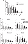
Distribution of PAI events in all subjects throughout the night. (A) Total numbers of each PAI event by intensity were summed per circadian hour starting at 10:00 PM and ending at 7:00 AM. Black symbols indicate PAI 15 events. Red symbols indicate PAI 18 events. Blue symbols indicate PAI 30 events. Green symbols indicate PAI 40 events. Yellow symbols indicate PAI 50 events. (B) Distribution of cortical arousal events in all subjects throughout the night.
Fig. 2.
Fig. 2.
Comparative amount of PAI events among good sleepers and poor sleepers throughout the night. Total numbers of each PAI were summed per hour for good sleepers and poor sleepers. (A) PAI 15. (B) PAI 18. (C) PAI 30. (D) PAI 40. (E) PAI 50. (F) Cortical arousals. , Good sleepers; ●, poor sleepers. *P < 0.05; **P < 0.01; ***P < 0.001.
Fig. 3.
Fig. 3.
Comparative nightly amounts of PAI and cortical arousals in good and poor sleepers. White bars indicate good sleepers, and black bars indicate poor sleepers. (A) Cumulative number of PAI events per night. (B) Number of PAI events per PSG-defined sleep state. (B, 1) Wake. (B, 2) LS: NREMS stages 1 and 2. (B, 3) DS: NREMS stage 3. (B, 4) REMS. *P < 0.05; **P < 0.01; ***P < 0.001.
Fig. 4.
Fig. 4.
Percentage of cortical arousals that had a PAI 40 or 50 event in close temporal proximity (±2 min). The first pair of bars indicates the percentage of events (cortical arousals or random times) that had a PAI 40/50 event in the 2-min period preceding it (Yes Before). The second pair of bars indicates the percentage of events (cortical arousals or random times) that had a PAI 40/50 event in the 2-min period following the event (Yes After). The third pair of bars indicates the percentage of events (cortical arousals or random times) that did not had a PAI 40/50 event in the 2-min period preceding it (No Before). The fourth pair of bars indicates the percentage of events (cortical arousals or random times) that did not had a PAI 40/50 event in the 2-min period following the event (No After). ***P < 0.001.
Fig. 5.
Fig. 5.
Dysregulation in poor sleepers in the number (A) and timing (B) of PAI events relative to a PSG-defined cortical arousal. (A) Total number of PAI 40 or 50 events in close temporal proximity to a PSG-defined cortical arousal. (B) Average time in seconds between PAI 40 or 50 and a PSG-defined cortical arousal. White bars indicate good sleepers, and black bars indicate poor sleepers. The first pair of bars is the total number of PAI 40 or 50 in the 2- to 1-min period preceding a PSG-defined cortical arousal. The second pair of bars is the total number of PAI 40 or 50 in the 1-min period preceding a PSG-defined cortical arousal. The third pair of bars is the total number of PAI 40 or 50 in the 1-min period following a PSG-defined cortical arousal. The fourth pair of bars is the total number of PAI 40 or 50 in the 1- to 2-min period following a PSG-defined cortical arousal. *P < 0.05; ***P < 0.001.
Fig. 6.
Fig. 6.
Temporal relationship between PAI 40 and 50 events and PSG-defined cortical arousals. The numbers of PAI 40 and 50 events in the 10-s epochs in the 1 min preceding and following a PSG-defined cortical arousal are shown. , Good sleepers; ●, poor sleepers. *P < 0.05; ***P < 0.001.

Similar articles

Cited by

References

    1. Pfaff D., Brain Arousal and Information Theory: Neural Genetic Mechanisms (Harvard University Press, 2006).
    1. Calderon D. P., Kilinc M., Maritan A., Banavar J. R., Pfaff D., Generalized CNS arousal: An elementary force within the vertebrate nervous system. Neurosci. Biobehav. Rev. 68, 167–176 (2016). - PMC - PubMed
    1. Saper C. B., Fuller P. M., Wake-sleep circuitry: An overview. Curr. Opin. Neurobiol. 44, 186–192 (2017). - PMC - PubMed
    1. Pfaff D., Ribeiro A., Matthews J., Kow L. M., Concepts and mechanisms of generalized central nervous system arousal. Ann. N. Y. Acad. Sci. 1129, 11–25 (2008). - PubMed
    1. Pfaff D. W., Martin E. M., Ribeiro A. C., Relations between mechanisms of CNS arousal and mechanisms of stress. Stress 10, 316–325 (2007). - PubMed

Publication types

MeSH terms