. 2022 Jun 21;119(25):e2122379119.
doi: 10.1073/pnas.2122379119. Epub 2022 Jun 13.

Memory-like NK cells armed with a neoepitope-specific CAR exhibit potent activity against NPM1 mutated acute myeloid leukemia

Affiliations

Memory-like NK cells armed with a neoepitope-specific CAR exhibit potent activity against NPM1 mutated acute myeloid leukemia

Han Dong et al. Proc Natl Acad Sci U S A. .

Abstract

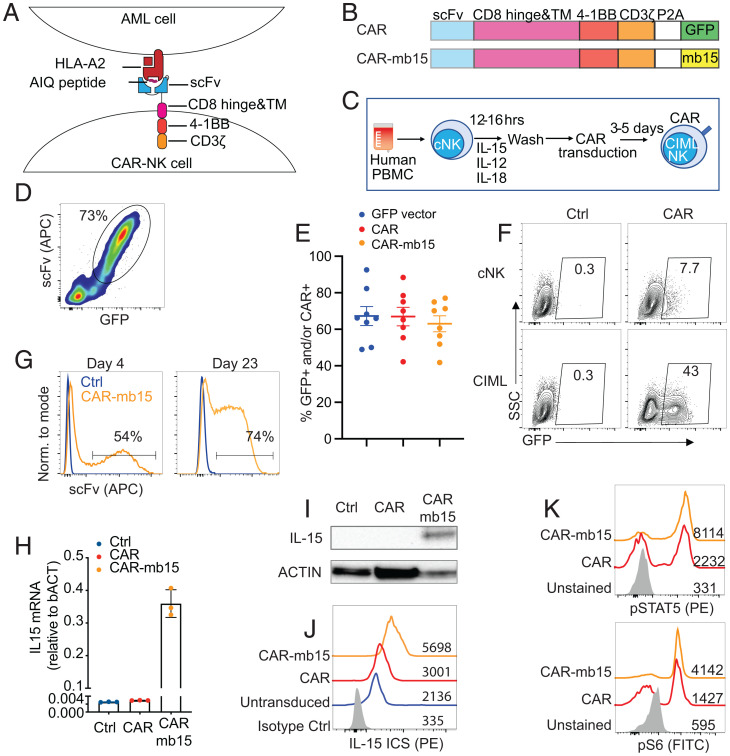
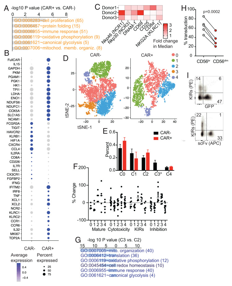
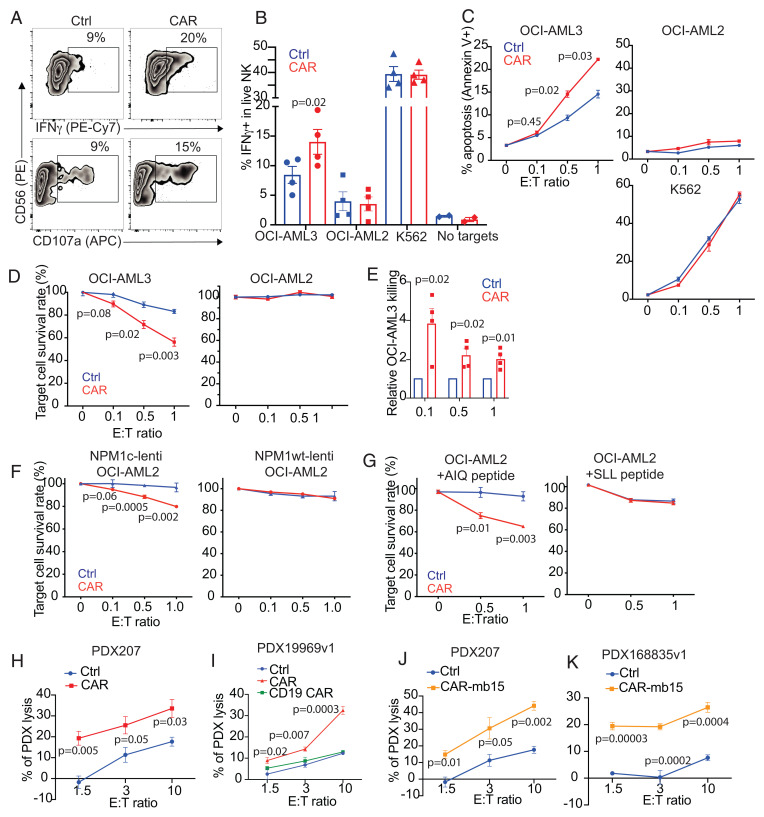
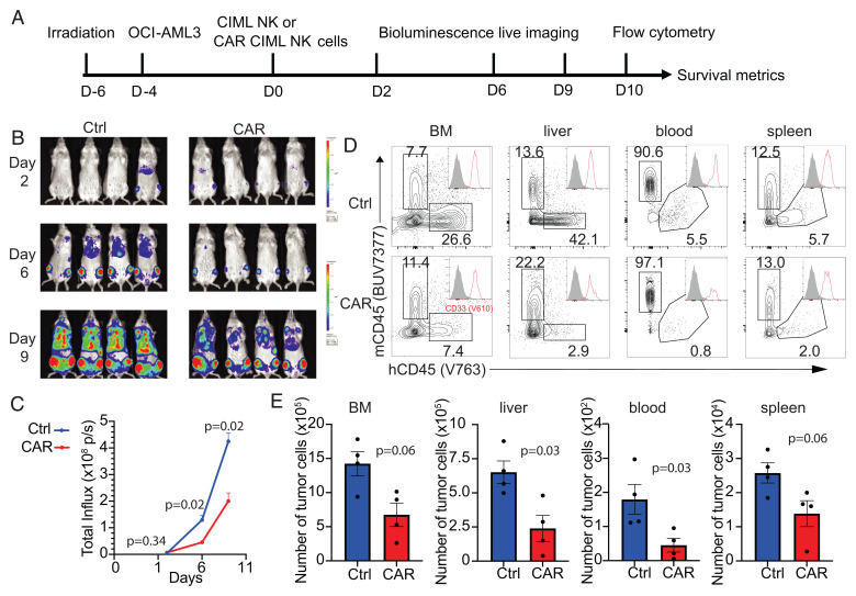
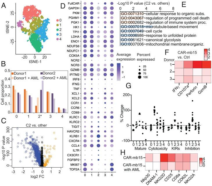
Acute myeloid leukemia (AML) remains a therapeutic challenge, and a paucity of tumor-specific targets has significantly hampered the development of effective immune-based therapies. Recent paradigm-changing studies have shown that natural killer (NK) cells exhibit innate memory upon brief activation with IL-12 and IL-18, leading to cytokine-induced memory-like (CIML) NK cell differentiation. CIML NK cells have enhanced antitumor activity and have shown promising results in early phase clinical trials in patients with relapsed/refractory AML. Here, we show that arming CIML NK cells with a neoepitope-specific chimeric antigen receptor (CAR) significantly enhances their antitumor responses to nucleophosphmin-1 (NPM1)-mutated AML while avoiding off-target toxicity. CIML NK cells differentiated from peripheral blood NK cells were efficiently transduced to express a TCR-like CAR that specifically recognizes a neoepitope derived from the cytosolic oncogenic NPM1-mutated protein presented by HLA-A2. These CAR CIML NK cells displayed enhanced activity against NPM1-mutated AML cell lines and patient-derived leukemic blast cells. CAR CIML NK cells persisted in vivo and significantly improved AML outcomes in xenograft models. Single-cell RNA sequencing and mass cytometry analyses identified up-regulation of cell proliferation, protein folding, immune responses, and major metabolic pathways in CAR-transduced CIML NK cells, resulting in tumor-specific, CAR-dependent activation and function in response to AML target cells. Thus, efficient arming of CIML NK cells with an NPM1-mutation-specific TCR-like CAR substantially improves their innate antitumor responses against an otherwise intracellular mutant protein. These preclinical findings justify evaluating this approach in clinical trials in HLA-A2+ AML patients with NPM1c mutations.

Keywords: CAR-NK cells; NPM1 mutation; TCR-like CAR; acute myeloid leukemia; memory-like NK cells.


Conflict of interest statement

Competing interest statement: R.R. has a sponsored research agreement with Crispr Therapeutics, Skyline Therapeutics and serves on the scientific advisory board of Glycostem Therapeutics. L.H.G. is a former Director of Bristol-Myers Squibb and the Waters Corporation and currently serves on the Board of Directors of GlaxoSmithKline Pharmaceuticals and Analog Devices, Inc. She also serves on the scientific advisory boards of Repare Therapeutics, Abpro Therapeutics and Kaleido Therapeutics. Other authors declare that they have no competing interests.

Figures

Fig. 1.
Fig. 1.
Generation of NPM1c-specific CAR-CIML NK cells. (A) Schematic diagram showing the design of CAR constructs and recognition of the AIQ/HLA-A2 complex on AML cells by NPM1c-CARs in B. (C) Schematic diagram showing the differentiation of freshly purified PB cNK cells into CIML NK cells and their transduction by BaEV-pseudotyped lentivirus. PBMC, peripheral blood mononuclear cell. (D) Representative flow cytometry plot showing expression of the NPM1c-CAR and GFP on transduced CIML NK cells. (E) Summary data showing transduction efficiencies of CIML NK cells from different donors with lentiviruses expressing GFP (blue dots), NPM1c-CAR (red dots), or NPM1c-CAR-mb15 (orange dots). (F) Comparison of transduction efficiency in cNK and CIML NK cells from a representative donor. Transduced cells were analyzed for GFP expression after 72 h, using untransduced cNK or CIML NK cells as controls (Ctrl). The numbers indicate percentages of cells in the gated area. (G) Representative flow cytometry histograms showing surface CAR expression of the untransduced (Ctrl in blue) and CAR-mb15-transduced (orange) CIML NK cells at day 4 and day 23 posttransduction. (H) qPCR quantification of IL15 transcripts and (I) Western blotting of IL-15 protein in CIML NK cells without transduction (Ctrl) or transduced with CAR or CAR-mb15. (J) Intracellular staining (ICS) of IL-15 in untransduced and CAR and CAR-mb15 transduced CIML NK cells by flow cytometry. Gray, blue, red, and orange histograms show isotype controls, untransduced, and CAR- and CAR-mb15 transduced CIML NK cells, respectively. (K) Flow cytometry assays for pSTAT5 (Upper Panel) and S6 kinase (Bottom Panel) in CAR and CAR-mb15 transduced CIML NK cells. Gray, red, and orange histograms show unstained controls and CAR- and CAR-mb15 transduced CIML NK cells, respectively. Data are representative of five (D) or three (F–K) independent experiments. Data in H show representative results using CIML NK cells from one PB donor, with error bars representing mean with SD from technical triplicates. Data in E are pooled from five independent experiments, with each dot representing one different PB donor; n = 8 for each column and error bars represented mean with SEM.
Fig. 2.
Fig. 2.
scRNA-seq and mass cytometry characterization of BaEV LV-transduced CAR-mb15 CIML NK cells. (A) GO functional enrichment analysis of DEGs between the CAR+ and CAR CIML NK cells (n = 2). DEGs were computed by FindMarkers between CAR+ and CAR populations with log2 fold-change of >0.1 and P < 0.05. (B) Dotplot of the expression of selected DEGs. (C) Heatmap of fold changes of selected NK cell activation and functional proteins between CAR+ and CAR CIML NK cells based on mass cytometry analysis. (D) tSNE clustering analysis of scRNA-seq data from CAR+ CIML NK cells (Right) and CAR CIML NK cells (Left). Each cluster between CAR+ and CAR cells was annotated based on the expression similarity of 2,000 variable genes. (E) Cell proportion of each cluster in CAR+ and CAR CIML NK cells. Note that cluster 3 was absent in CAR+ CIML NK cells. (F) The scores for maturation, cytotoxicity, KIR, and inhibition in each cluster. Each dot represents one gene in that cluster and category (SI Appendix, Materials and Methods). The score was calculated from the change of its expression to the average expression of all cells. (G) GO functional enrichment analysis of DEGs between cluster 3 and cluster 2 in CAR CIML NK cells. (H) Flow cytometry analysis of BaEV lentiviral transduction of CD56brigh (immature) versus CD56dim (mature) NK cells. (I) Representative flow cytometry plots showing negative correlation between KIR expression and CAR transduction with BaEV pseudotyped lentivirus expressing NPM1c-CAR and GFP. n = 3 (C), 2 (E), 5 (H) PB donors. Error bars in E represented mean with SD. Data in H were analyzed by two-tailed paired t test. Data are pooled from two (C) or three (H) independent experiments, or representative of two independent experiments (I).
Fig. 3.
Fig. 3.
NPM1c-CAR CIML NK cells exhibit potent and specific anti-AML function in vitro. (A) Representative flow cytometry staining profiles of IFNγ or CD107a versus CD56 of untransduced (Ctrl) and NPM1c-CAR transduced (CAR) CIML NK cells. Indicated NK cells were cocultured with OCI-AML3 target cells for 6 h and CD56+ cells were analyzed. (B) Summary data showing percentages of IFNγ-expressing cells in CAR-expressing vs. untransduced (Ctrl) CIML NK cells following incubation with OCI-AML3 (NPM1c+ HLA-A2+), OCI-AML2 (NPM1c HLA-A2+), K562 (HLA−), or no target cells. (C) Killing of OCI-AML3, OCI-AML2, and K562 target cells by untransduced (Ctrl) and CAR-transduced CIML NK cells at the indicated effector (E): target (T) ratios. CIML NK cells and target cells were incubated for 4 h, and percentages of annexin V+ tumor cells were assayed by flow cytometry. (D) Killing of OCI-AML3 and OCI-AML2 target cells by untransduced (Ctrl) and CAR-transduced CIML NK cells at the indicated E: T ratios. CIML NK cells and luciferase-expressing target cells were incubated for 24 h, and luciferase activity was quantified. (E) Summary data showing OCI-AML3 target killing by NPM1c-CAR CIML NK cells as assessed by luciferase assay. Data were normalized to the percentage of killing by the corresponding untransduced (Ctrl) CIML NK cells. (F) OCI-AML2 cells were transduced with lentivirus expressing NPM1c (Lenti-NPM1c, Left Panel) or the wild-type NPM1 control (Lenti-NPM1wt, Right Panel). Transduced cells were sorted, expanded, and used as target cells for killing assays as described in D. Killing of the transduced OCI-AML2 target cells by Ctrl and CAR-transduced CIML NK cells were measured at the indicated E:T ratios. (G) Comparison of killing of OCI-AML2 cells pulsed with 1 μM of AIQ (Left) or SLL (Right) peptides by NPM1c CAR-CIML NK cells or untransduced (Ctrl) CIML NK cells. NK cells were cocultured with peptide-pulsed OCI-AML2 target cells at the indicated E:T ratios for 24 h. Target cell killing was measured by the luciferase activity of surviving target cells. (H) Killing of low-passaged NPM1c+ HLA-A2+ PDX AML target cells by NPMc1c-CAR CIML NK cells in comparison with their untransduced CIML NK cells from the same PB donor (n = 1 PDX donor and 1 PB donor for each graph; error bars from technical triplicates). (I) Killing of NPM1c+ HLA-A2+ PDX AML target cells by untransduced (Ctrl), NPM1c CAR-CIML (CAR), and irrelevant CD19 CAR CIML NK cells. (J and K) Killing of NPM1c+ HLA-A2+ PDX target cells by NPMc1c-CAR-mb15 CIML NK cells (n = 2 PDX donors and 2 PB donors). Each dot in B and E represent one different PB donor. n = 4 in B and E. Error bars in B and E represented mean with SEM. Error bars in C, D, and F–K represented mean with SD from 3 to 5 technical replicates. Data were analyzed by two-tailed paired Student’s t test (B), two-tailed one sample t test (E), and two-tailed unpaired t test (C, D, and F–K). Data are pooled from two (B and E) independent experiments, or representative of three (C, D, F, and G) independent experiments.
Fig. 4.
Fig. 4.
NPM1c-CAR CIML NK cells are effective in controlling AML in vivo in a xenograft model. (A) Scheme of the experimental setup. NSG mice were sublethally irradiated, and 2 d later, luciferase+ OCI-AML3 cells (5 × 10⁵ cells) were injected IV. After 4 d, 1 × 10⁶ untransduced or transduced CIML NK cells were injected IV into the tumor-bearing mice. BLI was performed on the indicated days to monitor tumor burden. Mice were euthanized at 10 d post-NK cell adoptive transfer for flow cytometry analysis. (B and C) Comparison of tumor burden in recipient mice given untransduced (Ctrl) and transduced (CAR) CIML NK cells by BLI (B) and quantification of luciferase activity (C). (D) Representative flow cytometry staining profiles of hCD45 versus mCD45 gating on live cells in the indicated tissues. Inserts show CD33 histogram of hCD45+ cells. (E) Summary data showing absolute numbers of leukemic cells in the indicated tissues of recipient mice at day 10 after adoptive transfer with untransduced CIML (blue) vs. NPM1c CAR CIML NK cells (red) (n = 4 mice per group). Error bars in C and E represented mean with SEM. Data were analyzed by two-way ANOVA with the Sidak posttest (C) or two-tailed unpaired Student’s t test (E). Data are representative of three (B–E) independent experiments.
Fig. 5.
Fig. 5.
Incorporation of membrane-bound IL-15 (mb15) into the CAR design improves the efficacy of CAR CIML NK cells. (A) Representative flow cytometry plots showing cell viability (Upper Panel) and Ki-67 staining (Bottom Panel) of CAR- versus CAR-mb15-transduced CIML NK cells. CIML NK cells were cultured in cytokine-limited media for 5 d before flow cytometry assay. (B and C) Comparison of tumor burden in recipient mice given untransduced (Ctrl) or CAR- or CAR-mbIL15-transduced CIML NK cells by BLI (B) and quantification of luciferase activity (C) (n = 4 mice per group). (D–F) Comparison of AML burden and persistence of the transferred human NK cells in the indicated tissues of recipient mice treated with CIML NK cells expressing NPM1c-CAR or NPM1c-CAR-mb15 at day 19 post-NK cell adoptive transfer. (D) Representative flow cytometry staining profiles of hCD45 versus mCD45 gating on live cells (Left Panel) from liver, CD33 versus hCD45 gating on hCD45+ cells (Middle Panel), and CD56 versus anti-scFv gating on hCD45+ CD33 cells (Right Panel). (E) Quantifications of OCI-AML3 leukemic cells in the indicated tissues. (F) Quantifications of CAR-expressing human NK cell percentages in the indicated tissues. n = 4 mice per group. Error bars in C, E, and F represented mean with SEM from biological replicates. Data were analyzed by two-way ANOVA with the Sidak posttest (C) or one-way ANOVA with the Tukey posttest (E and F). Data are representative of two (A) or three (B–F) independent experiments.
Fig. 6.
Fig. 6.
scRNA-seq analyses reveal crosstalk between CAR-mb15 CIML NK cells and AML target cells. (A) tSNE clustering analysis of CAR+ NK cells cocultured with or without AML target cells. CIML NK cells generated from two PB donors were transduced with NPM1c-CAR-mb15 lentivirus, and purified viable NK cells were cocultured with OCI-AML3 cells for 24 h followed by scRNA-seq. (B) Cell proportion of identified clusters in CAR+ NK cells cocultured with or without AML target cells; cluster 2 was consistently increased in CAR+ NK cells cocultured with AML targets. (C) Volcano plot of gene expression between cluster 2 and other clusters. Blue dots: down-regulated; and orange dots: up-regulated DEGs. (D) Dot plots of the expression of selected genes. (E) GO functional enrichment analysis of DEGs in C. (F) Fold-change of IFNγ, CD107a, perforin, and granzyme B in NPM1c-CAR-mb15 CIML NK cells over untransduced (Ctrl) CIML NK cells. CIML NK cells from three different donors were cocultured with OCI-AML3 target cells for 5 h followed by mass cytometry analysis. (G) The scores for maturation, cytotoxicity, KIR, and inhibition in each cluster. (H) Comparison of selected marker expression by NPM1c-CAR-mb15 CIML NK cells and the untransduced CIML NK cells from the same donor. CAR+ and CAR CIML NK cells were cocultured with AML target cells for 5 h and analyzed by mass cytometry. The heatmap shows median values in protein expression levels of the indicated activation markers and receptors. n = 2 (A–E and G) and 3 (F) PB donors. Data are representative of two (H) independent experiments or pooled from two (F) independent experiments.

References

    1. Falini B., Tiacci E., Martelli M. P., Ascani S., Pileri S. A., New classification of acute myeloid leukemia and precursor-related neoplasms: Changes and unsolved issues. Discov. Med. 10, 281–292 (2010). - PubMed
    1. Venstrom J. M., et al. , HLA-C-dependent prevention of leukemia relapse by donor activating KIR2DS1. N. Engl. J. Med. 367, 805–816 (2012). - PMC - PubMed
    1. Cooley S., et al. , Donor selection for natural killer cell receptor genes leads to superior survival after unrelated transplantation for acute myelogenous leukemia. Blood 116, 2411–2419 (2010). - PMC - PubMed
    1. Bald T., Krummel M. F., Smyth M. J., Barry K. C., The NK cell-cancer cycle: Advances and new challenges in NK cell-based immunotherapies. Nat. Immunol. 21, 835–847 (2020). - PMC - PubMed
    1. Sivori S., et al. , NK cells and ILCs in tumor immunotherapy. Mol. Aspects Med. 80, 100870 (2021). - PubMed

Publication types

MeSH terms