. 2022 Jun 21;119(25):e2121867119.
doi: 10.1073/pnas.2121867119. Epub 2022 Jun 13.

Raf Kinase Inhibitory Protein regulates the cAMP-dependent protein kinase signaling pathway through a positive feedback loop

Affiliations

Raf Kinase Inhibitory Protein regulates the cAMP-dependent protein kinase signaling pathway through a positive feedback loop

Jiyoung Lee et al. Proc Natl Acad Sci U S A. .

Abstract

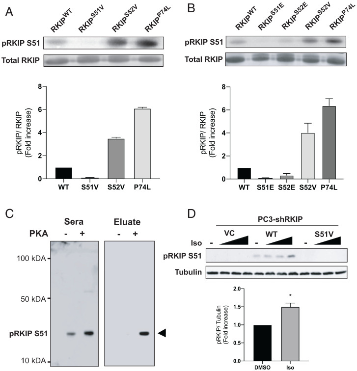
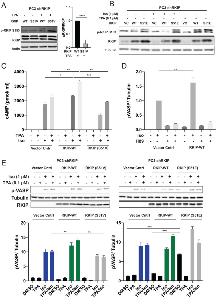
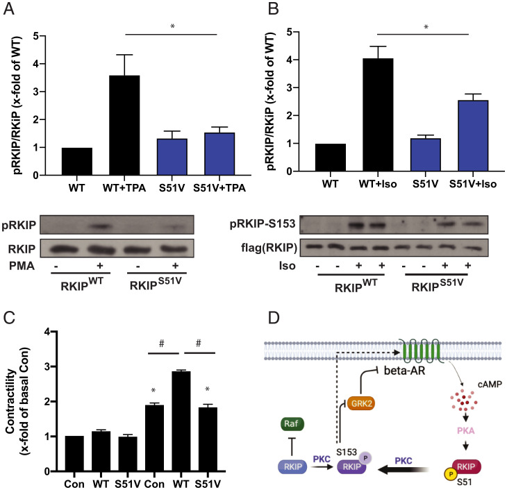
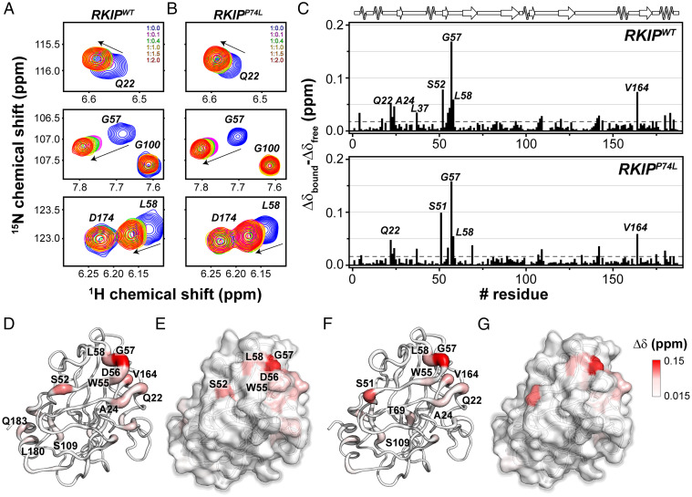
Raf Kinase Inhibitory Protein (RKIP) maintains cellular robustness and prevents the progression of diseases such as cancer and heart disease by regulating key kinase cascades including MAP kinase and protein kinase A (PKA). Phosphorylation of RKIP at S153 by Protein Kinase C (PKC) triggers a switch from inhibition of Raf to inhibition of the G protein coupled receptor kinase 2 (GRK2), enhancing signaling by the β-adrenergic receptor (β-AR) that activates PKA. Here we report that PKA-phosphorylated RKIP promotes β-AR-activated PKA signaling. Using biochemical, genetic, and biophysical approaches, we show that PKA phosphorylates RKIP at S51, increasing S153 phosphorylation by PKC and thereby triggering feedback activation of PKA. The S51V mutation blocks the ability of RKIP to activate PKA in prostate cancer cells and to induce contraction in primary cardiac myocytes in response to the β-AR activator isoproterenol, illustrating the functional importance of this positive feedback circuit. As previously shown for other kinases, phosphorylation of RKIP at S51 by PKA is enhanced upon RKIP destabilization by the P74L mutation. These results suggest that PKA phosphorylation at S51 may lead to allosteric changes associated with a higher-energy RKIP state that potentiates phosphorylation of RKIP at other key sites. This allosteric regulatory mechanism may have therapeutic potential for regulating PKA signaling in disease states.

Keywords: Raf Kinase Inhibitory Protein (RKIP); nuclear magnetic resonance (NMR); phosphatidylethanolamine binding protein (PEBP); protein kinase A (PKA).


Conflict of interest statement

The authors declare no competing interest.

Figures

Fig. 1.
Fig. 1.
Phosphorylation of RKIP by PKA. (A and B) Purified RKIP proteins were tested for in vitro kinase assays. After immunoblotting with anti-RKIP antibody, the phosphorylation levels were quantified using ImageQuant 5.2 software and normalized to RKIP protein levels to compare the phosphorylation between the various mutants. The fold induction shown in the bar graphs is an average of three experiments, and the error bars indicate SEM. (C) Immunoblotting purified WT (PKA-) and PKA-phosphorylated RKIP (PKA+) with unfractionated anti-pS51 antiserum (Sera) and purified antibody (Eluate). Rabbit serum was purified on an anti–phospho-S51 RKIP peptide column as described in Materials and Methods. (D) Immunoblotting of lysates from PC-3 prostate cancer cells treated with isoproterenol (Iso). PC-3 cells that had been stably depleted of RKIP by expression of human RKIP shRNA were transfected with a control vector (VC), RKIPWT, or the S51V RKIP mutant. Cells were lysed, and lysates were immunoblotted with anti-pS51 RKIP antibody or anti-tubulin antibody. For isoproterenol treatment, cells were serum-starved for 20 h and then either treated with DMSO or stimulated with 0.005, 0.05, or 1 μM isoproterenol (Iso) for 10 min. Cells were lysed, and lysates were immunoblotted with anti-pS51 RKIP antibody or anti-tubulin antibody. The protein bands were quantified using Licor ImageStudio, and the results were plotted for pS51 phosphorylation relative to tubulin. Mean values from four independent experiments were plotted, and P values were obtained using a Student’s t test. *P < 0.05.
Fig. 2.
Fig. 2.
PKA phosphorylates RKIP at Serine 51, increasing both PKC phosphorylation of RKIP at Serine 153 and PKA activity in prostate tumor cells. For AE, PC-3 cells stably expressing RKIP shRNA were transfected with control vector (VC), WT, or mutant RKIP (S51V, S51E) and serum-starved for 20 h before stimulation using TPA for 30 min, isoproterenol (Iso) for 10 min, or H89 for 30 min as indicated. (A) Phosphorylation of RKIP and mutant S51V RKIP at S153 in PC3 cells after TPA treatment. Cell lysates were immunoblotted with anti-pS153 RKIP antibody or anti-Actin antibody and quantified for plots. The lower band on phosphorylated RKIP and RKIP blots is nonspecific. (B) Phosphorylation of RKIP and mutant RKIP S51E at S513 after TPA and Iso treatment in PC3 cells. Cell lysates were immunoblotted with anti-pS153 RKIP antibody, anti-RKIP antibody, or anti-tubulin antibody. (C) Phosphorylation of S51 RKIP enhances cAMP levels in prostate tumor cells. Total cAMP levels were quantified as in Materials and Methods. (D) Phosphorylation of S51 RKIP enhances PKA activity in prostate tumor cells. Cell lysates were assayed for PKA activity by immunoblotting with anti-pVASP antibody. Samples were normalized to tubulin by immunoblotting with anti-tubulin antibody. (E) An S51V mutant reduces activation of PKA in TPA/Iso-stimulated cells, and S51E potentiates activation of PKA. Cell lysates were assayed for PKA activity by immunoblotting with anti-pVASP antibody. Samples were normalized to tubulin by immunoblotting with anti-tubulin antibody. Samples were quantified using Licor ImageStudio. Mean values from three independent experiments were plotted, and P values were obtained using a Student’s t test. *P < 0.05, **P < 0.01, ***P < 0.001, ****P < 0.0001.
Fig. 3.
Fig. 3.
Positive feedback loop between RKIP and PKA leads to enhanced contractility in cardiac myocytes. (A) Cardiac myocytes were adenovirally transduced with WT or S51V RKIP, and treated with or without TPA (PMA). Cell lysates were immunoblotted for RKIP or pS153 RKIP. (B) Cardiac myocytes were adenovirally transduced with WT or S51V RKIP and then treated with or without isoproterenol. Cell lysates were immunoblotted for RKIP or pS153 RKIP. (C) Cardiac myocytes were adenovirally transduced and treated with isoproterenol as in B. For the control (Con), LacZ was adenovirally transduced. Contractility was measured as described in Materials and Methods. Mean values from five (A) or six (B) independent experiments were plotted, and P values were obtained using a one-way ANOVA and Sidak’s post hoc test (WT + PMA vs. V51 + PMA: P = 0.0178 and WT + ISO vs. V51 + ISO: P = 0.0016). (C) Mean values from 20 replicates of five independent experiments were plotted, and P values were obtained using a one-way ANOVA and Tukey’s post hoc test (*P < 0.01 vs. Con-Iso and #P < 0.0001 as indicated). (D) Scheme depicting positive feedback loop between RKIP and PKA. RKIP phosphorylated at S153 by PKC inhibits GRK2, reducing down-regulation of the β-adrenergic receptor (β-AR). β-AR activates PKA, which then phosphorylates RKIP at S51 leading to enhanced phosphorylation by PKC at S153.
Fig. 4.
Fig. 4.
NMR map of the residues of RKIPWT and P74L mutant involved in PKA-C/RKIP interaction interface. (A and B) [1H,15N]-HSQC spectra showing the backbone chemical shift changes of selected resonances of (A) RKIPWT and (B) RKIPP74L upon titration of increasing amount of PKA-C/ATPγN complex. (C) CSP of the amide fingerprint of RKIPWT and RKIPP74L mutant alone (Δδfree) and in a 1:2 complex with PKA-C/ATPγN (Δδbound) vs. residues, calculated using Eq. 1. The residues that show a CSP value two times greater than 1 SD from the average CSP (gray dashed line) are indicated in the histogram. (DG) Cartoon (D) and surface (E) mapping of the amide CSP of RKIPWT or cartoon (F) and surface (G) of P74L mutant onto the three-dimensional structure (Protein Data Bank [PDB] ID 2IQY). The cartoons and the surfaces are colored according the CSP values ranging from 0.015 to 0.15 ppm.
Fig. 5.
Fig. 5.
NMR mapping of the PKA-C residues involved in the PKA-C/RKIP interactions. (A) CSPs of the PKA-C/ATPγN fingerprint upon interaction with RKIPWT (black) and PKI. The shaded colors indicate the Gly-rich loop (green), catalytic loop (yellow), DFG loop (orange), activation loop (purple), P + 1 loop (blue), and APE (red). (B) Chemical shift changes mapped onto the PKA-C structure (PDB ID 4wb5). The hot pink spheres highlight residues that broaden out, while the orange spheres indicate those residues that sharpened out upon interacting with RKIPWT. (C) [1H,15N]-TROSY-HSQC spectra overlay for specific residues of PKA-C in complex with ATPγN (orange), ATPγN/RKIP (purple), and ATPγN/PKI (green).

Similar articles

Cited by

References

    1. Brandman O., Ferrell J. E. Jr., Li R., Meyer T., Interlinked fast and slow positive feedback loops drive reliable cell decisions. Science 310, 496–498 (2005). - PMC - PubMed
    1. Lorenz K., Rosner M. R., Brand T., Schmitt J. P., Raf kinase inhibitor protein: Lessons of a better way for β-adrenergic receptor activation in the heart. J. Physiol. 595, 4073–4087 (2017). - PMC - PubMed
    1. Yesilkanal A. E., Rosner M. R., Targeting Raf kinase inhibitory protein regulation and function. Cancers (Basel) 10, E306 (2018). - PMC - PubMed
    1. Dangi-Garimella S., et al. , Raf kinase inhibitory protein suppresses a metastasis signalling cascade involving LIN28 and let-7. EMBO J. 28, 347–358 (2009). - PMC - PubMed
    1. Yun J., et al. , Signalling pathway for RKIP and Let-7 regulates and predicts metastatic breast cancer. EMBO J. 30, 4500–4514 (2011). - PMC - PubMed

Publication types

MeSH terms

Substances