. 2022 Jun 13;12(6):e059110.
doi: 10.1136/bmjopen-2021-059110.

Chronic lung lesions in COVID-19 survivors: predictive clinical model

Collaborators, Affiliations

Chronic lung lesions in COVID-19 survivors: predictive clinical model

Carlos Roberto Ribeiro Carvalho et al. BMJ Open. .

Abstract

Objective: This study aimed to propose a simple, accessible and low-cost predictive clinical model to detect lung lesions due to COVID-19 infection.

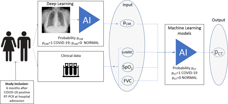
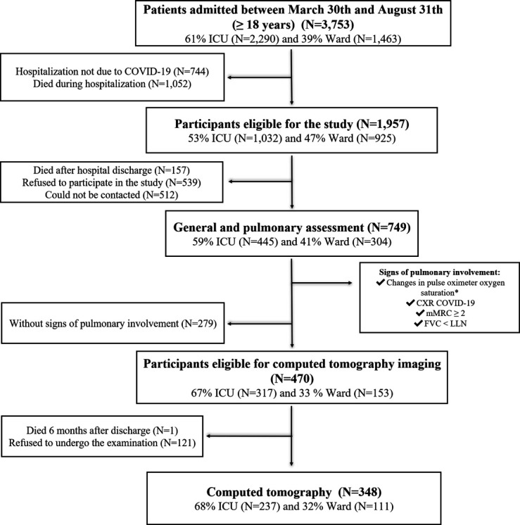
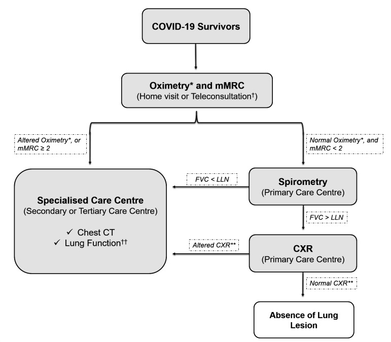
Design: This prospective cohort study included COVID-19 survivors hospitalised between 30 March 2020 and 31 August 2020 followed-up 6 months after hospital discharge. The pulmonary function was assessed using the modified Medical Research Council (mMRC) dyspnoea scale, oximetry (SpO2), spirometry (forced vital capacity (FVC)) and chest X-ray (CXR) during an in-person consultation. Patients with abnormalities in at least one of these parameters underwent chest CT. mMRC scale, SpO2, FVC and CXR findings were used to build a machine learning model for lung lesion detection on CT.

Setting: A tertiary hospital in Sao Paulo, Brazil.

Participants: 749 eligible RT-PCR-confirmed SARS-CoV-2-infected patients aged ≥18 years.

Primary outcome measure: A predictive clinical model for lung lesion detection on chest CT.

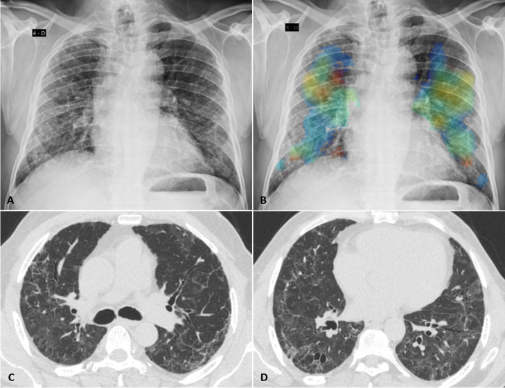
Results: There were 470 patients (63%) that had at least one sign of pulmonary involvement and were eligible for CT. Almost half of them (48%) had significant pulmonary abnormalities, including ground-glass opacities, parenchymal bands, reticulation, traction bronchiectasis and architectural distortion. The machine learning model, including the results of 257 patients with complete data on mMRC, SpO2, FVC, CXR and CT, accurately detected pulmonary lesions by the joint data of CXR, mMRC scale, SpO2 and FVC (sensitivity, 0.85±0.08; specificity, 0.70±0.06; F1-score, 0.79±0.06 and area under the curve, 0.80±0.07).

Conclusion: A predictive clinical model based on CXR, mMRC, oximetry and spirometry data can accurately screen patients with lung lesions after SARS-CoV-2 infection. Given that these examinations are highly accessible and low cost, this protocol can be automated and implemented in different countries for early detection of COVID-19 sequelae.

Keywords: COVID-19; chest imaging; respiratory medicine (see thoracic medicine).


Conflict of interest statement

Competing interests: None declared.

Figures

Figure 1
Figure 1
Logistic regression-based machine learning model to detect the presence of COVID-19-related lung lesions. The patients were invited to participate in the study 6 months after COVID-19-positive RT-PCR at hospital admission. The modified Medical Research Council (mMRC) dyspnoea scale, oximetry (SpO2), spirometry (forced vital capacity (FVC)) and the five radiographic scores obtained during DL-based classification of chest X-ray (pCXR) were used as input data, and the presence of lung lesions due to COVID-19 was used as output data. AI, artificial intelligence.
Figure 2
Figure 2
Flow chart of patient selection. *Rest SpO2 <90% or a decrease in SpO2 of at least 4% after the 1 min sit and stand test. CXR, chest X-ray; FVC, forced vital capacity; ICU, intensive care unit; LLN, lower limit of normal; mMRC, modified Medical Research Council dyspnoea scale.
Figure 3
Figure 3
Fibrotic-like changes after critical COVID-19 in a patient in his early 70s. (A) Posteroanterior chest radiograph obtained 7 months after infection shows reticular opacities with a slight peripheral predominance diffusely distributed in both lungs. (B) Image from the same radiograph analysed by the artificial intelligence algorithm with a heat map highlighting the areas of pulmonary involvement. (C, D) Chest CT obtained 8 months after infection shows moderate ground glass opacities, linear multifocal and reticular abnormalities, discrete traction bronchiectasis and slight parenchymal architectural distortion. The patient had dyspnoea (modified Medical Research Council dyspnoea scale=1) and altered forced vital capacity (2.34 L/60% pred), besides the normal oximetry (97%).
Figure 4
Figure 4
Flow chart for lung lesion case-finding in COVID-19 survivors. *Altered oximetry: resting SpO2 ≤90% or a decrease in SpO2 of ≥4% during the 1 min sit and stand test. **Altered CXR: COVID-19 findings, including bilateral linear and/or reticular opacities, especially peripheral opacities. †The in-person consultation also should start with oximetry and mMRC examinations. ††The suggestion is to perform plethysmography with diffusion capacity measure. CXR, chest X-ray; FVC, forced vital capacity; LLN, lower limit of normal; mMRC, modified Medical Research Council dyspnoea scale.

References

    1. Sisó-Almirall A, Brito-Zerón P, Conangla Ferrín L, et al. . Long Covid-19: proposed primary care clinical guidelines for diagnosis and disease management. Int J Environ Res Public Health 2021;18. 10.3390/ijerph18084350. [Epub ahead of print: 20 04 2021]. - DOI - PMC - PubMed
    1. Nalbandian A, Sehgal K, Gupta A, et al. . Post-acute COVID-19 syndrome. Nat Med 2021;27:601–15. 10.1038/s41591-021-01283-z - DOI - PMC - PubMed
    1. Mauad T, Duarte-Neto AN, da Silva LFF, et al. . Tracking the time course of pathological patterns of lung injury in severe COVID-19. Respir Res 2021;22:32. 10.1186/s12931-021-01628-9 - DOI - PMC - PubMed
    1. Tanni SE, Fabro AT, de Albuquerque A, et al. . Pulmonary fibrosis secondary to COVID-19: a narrative review. Expert Rev Respir Med 2021;15:791–803. 10.1080/17476348.2021.1916472 - DOI - PubMed
    1. Macedo BRde, Garcia MVF, Garcia ML, et al. . Implementation of Tele-ICU during the COVID-19 pandemic. J Bras Pneumol 2021;47:e20200545. 10.36416/1806-3756/e20200545 - DOI - PMC - PubMed

Publication types