Review
. 2022 Jun 13;93(9):919-929.
doi: 10.1136/jnnp-2021-328586. Online ahead of print.

Neuropathology and emerging biomarkers in corticobasal syndrome

Affiliations
Review

Neuropathology and emerging biomarkers in corticobasal syndrome

Shunsuke Koga et al. J Neurol Neurosurg Psychiatry. .

Abstract

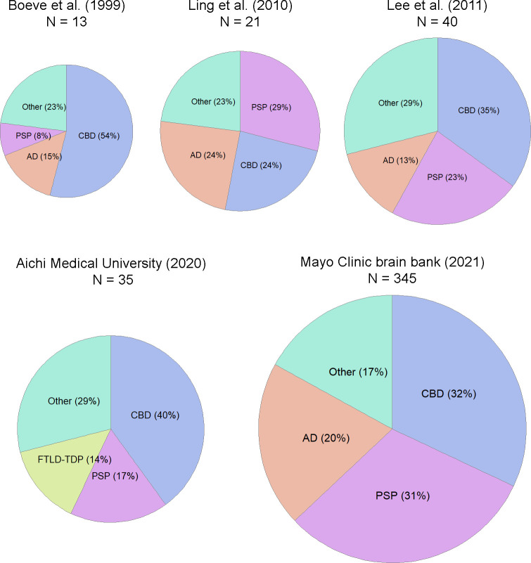
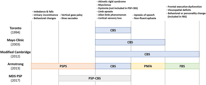
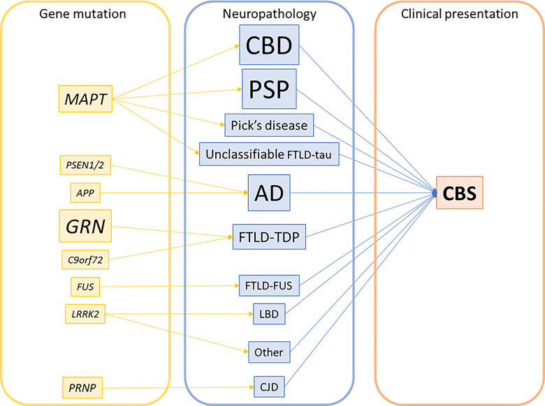
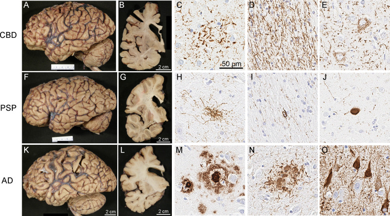
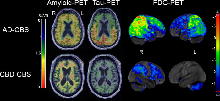
Corticobasal syndrome (CBS) is a clinical syndrome characterised by progressive asymmetric limb rigidity and apraxia with dystonia, myoclonus, cortical sensory loss and alien limb phenomenon. Corticobasal degeneration (CBD) is one of the most common underlying pathologies of CBS, but other disorders, such as progressive supranuclear palsy (PSP), Alzheimer's disease (AD) and frontotemporal lobar degeneration with TDP-43 inclusions, are also associated with this syndrome.In this review, we describe common and rare neuropathological findings in CBS, including tauopathies, synucleinopathies, TDP-43 proteinopathies, fused in sarcoma proteinopathy, prion disease (Creutzfeldt-Jakob disease) and cerebrovascular disease, based on a narrative review of the literature and clinicopathological studies from two brain banks. Genetic mutations associated with CBS, including GRN and MAPT, are also reviewed. Clinicopathological studies on neurodegenerative disorders associated with CBS have shown that regardless of the underlying pathology, frontoparietal, as well as motor and premotor pathology is associated with CBS. Clinical features that can predict the underlying pathology of CBS remain unclear. Using AD-related biomarkers (ie, amyloid and tau positron emission tomography (PET) and fluid biomarkers), CBS caused by AD often can be differentiated from other causes of CBS. Tau PET may help distinguish AD from other tauopathies and non-tauopathies, but it remains challenging to differentiate non-AD tauopathies, especially PSP and CBD. Although the current clinical diagnostic criteria for CBS have suboptimal sensitivity and specificity, emerging biomarkers hold promise for future improvements in the diagnosis of underlying pathology in patients with CBS.

Keywords: alzheimer's disease; corticobasal degeneration; genetics; neuropathology; supranuclear palsy.


Conflict of interest statement

Competing interests: None declared.

Figures

Figure 1
Figure 1
Frequency of the common underlying pathologies associated with CBS. The pie charts (not drawn to scale) on the upper row are from review of the literature. The pie charts on the lower row are from unpublished data from the brain bank at the Institute for Medical Science of Aging, Aichi Medical University (1994–2020) and the Mayo Clinic brain bank (1998–2021). CBD is the most frequent underlying pathology, followed by PSP and AD. AD, Alzheimer’s disease; CBD, corticobasal degeneration; CBS, corticobasal syndrome; FTLD-TDP, frontotemporal lobar degeneration with TDP-43 inclusions; PSP, progressive supranuclear palsy.
Figure 2
Figure 2
Comparison of diagnostic criteria for CBS. Five commonly used criteria for CBS are compared. The motor symptoms and signs are in common in CBS, but the cognitive and language impairment are differently included between Mayo Clinic criteria, University of Toronto criteria and the modified Cambridge criteria. Armstrong criteria define four clinical presentations in CBD. The CBS of the modified Cambridge criteria roughly corresponds to CBS, PNFA and FBS of the Armstrong criteria. The MDS diagnostic criteria for PSP define PSP-CBS, which partially overlaps PSP syndrome of Armstrong criteria, although dystonia is not included in the criteria for PSP-CBS. Both Armstrong and MDS criteria list exclusion criteria, which suggest other underlying pathology. CBS, corticobasal syndrome; FBS, frontal behavioral-spatial syndrome; MDS, International Parkinson and Movement Disorder Society; PNFA, progressive non-fluent aphasia; PSPS, progressive supranuclear palsy syndrome.
Figure 3
Figure 3
Genetic–pathological–clinical correlations in CBS. Font size reflects the frequency of each mutation or pathological diagnosis. Note that the majority of pathologies are sporadic; only a small subset of cases has gene mutations indicated. AD, Alzheimer’s disease; CBD, corticobasal degeneration; CBS, corticobasal syndrome; CJD, Creutzfeldt-Jakob disease; FTLD-FUS, frontotemporal lobar degeneration with fused-in-sarcoma pathology; FTLD-TDP, frontotemporal lobar degeneration with TDP-43 inclusions; LBD, Lewy body disease; PSP, progressive supranuclear palsy.
Figure 4
Figure 4
Macroscopic and microscopic findings of patients with CBS and underlying pathology of CBD (A–E), PSP (F–J), and AD (K–O). Microscopic images show immunohistochemistry for phosphorylated-tau (C–E, H–J, N–O) and amyloid-β (panel M). In CBD, the characteristic findings include astrocytic plaques (C), numerous tau-positive threads in the white matter (D) and pretangles (E). In PSP, the characteristic findings are tufted astrocytes (H), oligodendroglial coiled bodies (I), and globose neurofibrillary tangles (J). In AD, the characteristic findings with tau immunohistochemistry are neuritic plaques (N) and neurofibrillary tangles (O), as well as amyloid-β positive amyloid plaques (M). Gross patterns of brain atrophy (A, B, F, G, K, L) are not specific, but show frontal lobe atrophy and enlargement of the frontal horn of the lateral ventricle in cases with CBS. AD, Alzheimer’s disease; CBD, corticobasal degeneration; PSP, progressive supranuclear palsy.
Figure 5
Figure 5
Representative amyloid PET with [11C]PiB, tau PET with [18F]flortaucipir and glucose PET with [18F]fluoro-2-deoxyglucose (FDG) in AD-CBS and CBD-CBS. The amyloid PET and tau PET are shown on representative axial slices on the same colour scale, while the FDG-PET highlights regional metabolism with z-scores between −1 and −7 SD from controls using CortexID suite software. Upper images are from a patient with AD-CBS. Amyloid PET shows diffuse ligand uptake in cortical grey matter, and tau PET reveals increased signal in the temporo-parieto-occipital lobes. FDG-PET reveals hypometabolism in bilateral parietal cortex extending into occipital lobes (right >left) and right >left superior frontal cortex. Lower images are from a patient with CBD-CBS. There is no evidence of increase amyloid PET uptake in the cortical grey matter and there is no increase tau PET uptake. FDG-PET reveals mild hypometabolism in the right premotor cortex and postcentral gyrus in CBD-CBS. AD-CBS, Alzheimer’s disease presenting as corticobasal syndrome; CBD-CBS, corticobasal degeneration presenting as corticobasal syndrome; FDG, fluorodeoxyglucose; L, left; PET, positron emission tomography; PIB, Pittsburgh compound-B; R, right; SUVR, standardised uptake value ratio.

Comment in

References

    1. Rebeiz JJ, Kolodny EH, Richardson EP. Corticodentatonigral degeneration with neuronal achromasia. Arch Neurol 1968;18:20–33. 10.1001/archneur.1968.00470310034003 - DOI - PubMed
    1. Boeve BF, Maraganore DM, Parisi JE, et al. . Pathologic heterogeneity in clinically diagnosed corticobasal degeneration. Neurology 1999;53:795–800. 10.1212/wnl.53.4.795 - DOI - PubMed
    1. Cordato NJ, Halliday GM, McCann H, et al. . Corticobasal syndrome with tau pathology. Mov Disord 2001;16:656–67. 10.1002/mds.1124 - DOI - PubMed
    1. Boeve BF, Lang AE, Litvan I. Corticobasal degeneration and its relationship to progressive supranuclear palsy and frontotemporal dementia. Ann Neurol 2003;54 Suppl 5:S15–19. 10.1002/ana.10570 - DOI - PubMed
    1. Lang A, Riley DE, Bergeron C. Cortical-Basal ganglionic degeneration. neurodegenerative diseases. Philadelphia: WB Saunders, 1994: 877–94.