Review
. 2022 Jun 13;7(1):186.
doi: 10.1038/s41392-022-01043-6.

Cell deaths: Involvement in the pathogenesis and intervention therapy of COVID-19

Affiliations
Review

Cell deaths: Involvement in the pathogenesis and intervention therapy of COVID-19

Xue Li et al. Signal Transduct Target Ther. .

Abstract

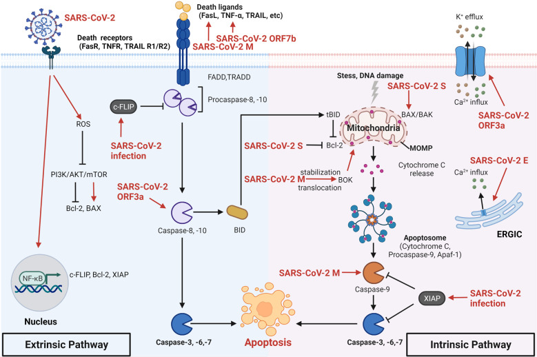
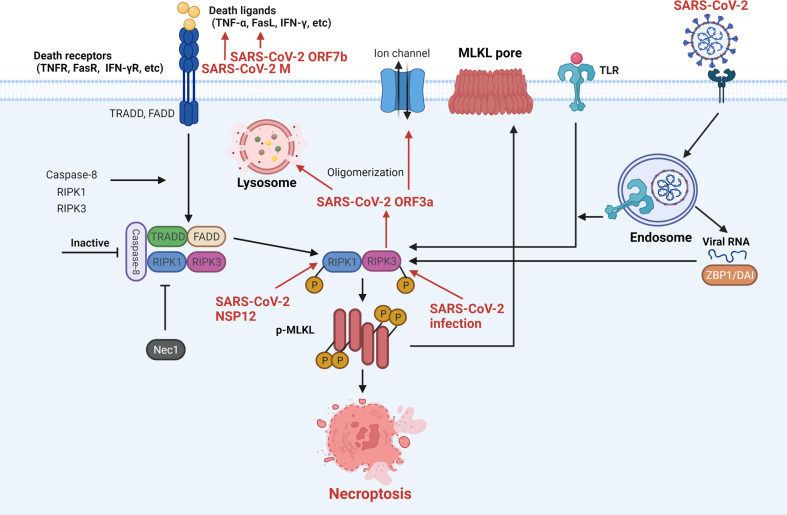
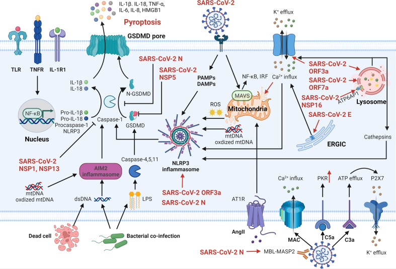
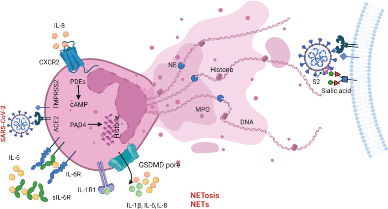
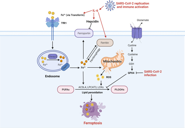
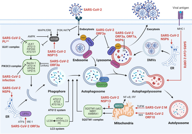
The current pandemic of coronavirus disease 2019 (COVID-19) caused by severe acute respiratory syndrome coronavirus 2 (SARS-CoV-2) infection has dramatically influenced various aspects of the world. It is urgent to thoroughly study pathology and underlying mechanisms for developing effective strategies to prevent and treat this threatening disease. It is universally acknowledged that cell death and cell autophagy are essential and crucial to maintaining host homeostasis and participating in disease pathogenesis. At present, more than twenty different types of cell death have been discovered, some parts of which have been fully understood, whereas some of which need more investigation. Increasing studies have indicated that cell death and cell autophagy caused by coronavirus might play an important role in virus infection and pathogenicity. However, the knowledge of the interactions and related mechanisms of SARS-CoV-2 between cell death and cell autophagy lacks systematic elucidation. Therefore, in this review, we comprehensively delineate how SARS-CoV-2 manipulates diverse cell death (including apoptosis, necroptosis, pyroptosis, ferroptosis, and NETosis) and cell autophagy for itself benefits, which is simultaneously involved in the occurrence and progression of COVID-19, aiming to provide a reasonable basis for the existing interventions and further development of novel therapies.


Conflict of interest statement

The authors declare no competing interests.

Figures

Fig. 1
Fig. 1
The mechanisms of SARS-CoV-2 regulating cell apoptosis. Activated death receptors recruit and cleave caspase-8/10, subsequently activating caspase-3/6/7 to induce extrinsic cell apoptosis. Stress- and DNA damage-stimulated cytochrome c directly cleaves caspase-9 to activate caspase-3/6/7 for intrinsic cell apoptosis. And tBID caused by caspase-8 also induces intrinsic apoptotic way. SARS-CoV-2 infection-upregulated c-FLIP and NF-κB inhibit apoptosis in infected cells. In contrast, SARS-CoV-2 ORF7b- and M protein-upregulated death ligands and SARS-CoV-2 ORF3a-activated caspase-8 promote extrinsic apoptosis. And SARS-CoV-2 S protein regulates Bcl-2 and BAX to induce the intrinsic apoptotic pathway. SARS-CoV-2 M protein-stabilized BOK and -directly activated caspase-9 both induce intrinsic apoptosis. Besides, cellular ion imbalance caused by SARS-CoV-2 ORF3a and E protein is involved in infected cell apoptosis. SARS-CoV-2 severe acute respiratory syndrome coronavirus 2, S spike, E envelope, M membrane, ORF open reading frame, NF-κB nuclear factor kappa B, c-FLIP cellular Fas-associated protein with death domain-like interleukin-1-converting enzyme-inhibitory protein, Bcl-2 B-cell lymphoma-2, XIAP X-linked inhibitor of apoptosis protein, ROS reactive oxygen species, PI3K phosphoinositide 3-kinase, AKT protein kinase B, mTOR mammalian target of rapamycin, TNF-α tumor necrosis factor-alpha, TRAIL TNF-related apoptosis-inducing ligand, FADD Fas-associated protein with death domain, TRADD TNF receptor 1-associated death domain protein, Caspase cysteine-aspartic protease, tBID truncated BH3 interacting domain death agonist, BOK Bcl-2 ovarian killer, BAX Bcl-2-associated X, BAK Bcl-2 homologous killer, MOMP mitochondrial outer membrane permeabilization, Apaf-1 apoptotic protease activating factor-1, ERGIC endoplasmic reticulum-Golgi intermediate compartment. Created with BioRender
Fig. 2
Fig. 2
The interaction of SARS-CoV-2 and cell necroptosis. Activated death receptors phosphorylate RIPK1 and RIPK3 in the absence of active caspase-8, and consecutively phosphorylate MLKL to form pores in the cell membrane, eventually leading to cell necroptosis. SARS-CoV-2 ORF7b and M protein-upregulated death ligands, SARS-CoV-2 NSP12-activated RIPK1, and SARS-CoV-2-related RIPK3 activation all promote necroptosis in infected cells. Meanwhile, the SARS-CoV-2-stimulated TLRs pathway and viral ZBP1 axis also directly activate RIPK3 to induce necroptosis. Besides, RIPK3-induced SARS-CoV-2 ORF3a oligomerization enhances its functions. SARS-CoV-2 severe acute respiratory syndrome coronavirus 2, M membrane, ORF open reading frame, NSP nonstructural protein, IFN-γ interferon-gamma, TRADD TNF receptor 1-associated death domain protein, FADD Fas-associated protein with death domain, Caspase cysteine-aspartic protease, RIPK receptor-interacting protein kinase, Nec1 necrostatin-1, MLKL mixed lineage kinase domain-like protein, TLR Toll-like receptor, ZBP1/DAI Z-DNA/RNA binding protein-1. Created with BioRender
Fig. 3
Fig. 3
The sophisticated associations of SARS-CoV-2, host system, and cell pyroptosis. Activated TLRs, TNFR, and IL-1R1 pathways induce NF-κB activation to promote pro-inflammatory cytokines production including pro-IL-1β, pro-IL-18, procaspase-1, and NLRP3. The PAMPs and DAMPs caused by SARS-CoV-2 immediately activate the NLRP3 inflammasome to cleave procaspase-1 that subsequently cleaves pro-IL-1β and pro-IL-18. Meanwhile, caspase-1-cleaved GSDMD inserts into the membrane to form pores, leading to cell pyroptosis and contents release. SARS-CoV-2 N protein, NSP5, NSP1, and NSP13 inhibit infected cell pyroptosis via impeding the caspase-1 or GSDMD. On the contrary, more commonly, SARS-CoV-2 promotes cell pyroptosis in various ways. SARS-CoV-2 ORF3a and N protein directly activate the NLRP3 inflammasome. The ion imbalance caused by SARS-CoV-2 ORF3a and E protein and lysosomal dysfunction caused by SARS-CoV-2 ORF3a, ORF7a, and NSP16 both promote the NLRP3 inflammasome activation. In addition, the MAVS pathway activated by SARS-CoV-2 infection causes mitochondria damage and the activation of NF-κB and IRF. Besides, SARS-CoV-2-related ROS production and mtDNA release also activate the NLRP3 inflammasome. Meanwhile, dsDNA and mtDNA-induced AIM2 inflammasome and LPS-induced caspase-4/5/11 also induce cell pyroptosis. From the perspective of the host, the Ang II-AT1R axis and the MBL-MASP-mediated complement system are involved in infected cell pyroptosis. SARS-CoV-2 severe acute respiratory syndrome coronavirus 2, N nucleocapsid, E envelope, ORF open reading frame, NSP nonstructural protein, TLR Toll-like receptor, TNFR tumor necrosis factor receptor, IL-1R1 interleukin-1 receptor 1, NF-κB nuclear factor kappa B, IRF transcription factors interferon regulatory factor, TNF-α tumor necrosis factor-alpha, HMGB1 high mobility group box 1, GSDMD gasdermin-D, Caspase cysteine-aspartic protease, AIM2 absent in melanoma, dsDNA double-stranded DNA, LPS lipopolysaccharide, PAMPs pathogen-associated molecular patterns, DAMPs damage-associated molecular patterns, NLRP3 nucleotide-binding oligomerization domain-like receptor containing pyrin domain 3, ROS reactive oxygen species, mtDNA mitochondrial DNA, Ang II angiotensin II, AT1R Ang II-angiotensin 1 receptor, MBL-MASP mannan-binding lectin (MBL)-MBL-associated serine protease, MAC membrane attack complex, PKR double-stranded RNA-dependent protein kinase, ERGIC endoplasmic reticulum-Golgi intermediate compartment. Created with BioRender
Fig. 4
Fig. 4. The NETosis during SARS-CoV-2 infection.
NETs, released by SARS-CoV-2-induced NETosis, are comprised of DNA, histones, MPO, and NE. The IL-8-CXCR2, IL-6-IL-6R, and IL-1β-IL-1R1 axis play important roles in recruiting and activating neutrophils. In addition, SARS-CoV-2 hijacks histones to interact with S2 and sialic acid for enhanced infection. SARS-CoV-2 severe acute respiratory syndrome coronavirus 2, S2 subunit 2 of SARS-CoV-2 spike protein, ACE2 angiotensin-converting enzyme-2, TMPRSS2 transmembrane serine protease 2, IL-8 interleukin-8, CXCR2 C-X-C motif chemokine receptor 2, sIl-6R soluble IL-6 receptor, IL-1R1 interleukin-1 receptor 1, GSDMD gasdermin-D, PDEs phosphodiesterases, cAMP cyclic adenosine monophosphate, PAD4 peptidylarginine deiminase 4, NE neutrophil elastase, MPO myeloperoxidase, NETs neutrophil extracellular traps. Created with BioRender
Fig. 5
Fig. 5
The ferroptosis upon SARS-CoV-2 infection. Ferroptosis is triggered by lipid peroxidation resulting from the imbalance among iron metabolism, ROS generation, and antioxidant system (especially the cystine-GSH-GPX4 axis). The high level of IL-6 in COVID-19 directly promotes the expression of iron accumulation proteins (including transferrin and ferritin) and indirectly inhibits the exported protein ferroportin via hepcidin, leading to cell ferroptosis. Meanwhile, SARS-CoV-2 infection exhibits a negative effect on GPX4. SARS-CoV-2 severe acute respiratory syndrome coronavirus 2, IL-6 interleukin-6, TfR1 transferrin receptor 1, Steap3 six-transmembrane epithelial antigen of prostate 3, DMT1 divalent metal transporter 1, ROS reactive oxygen species, PUFAs polyunsaturated fatty acids, PLOOHs phospholipid hydroperoxides, ACSL4 acyl-CoA synthetase long-chain family member 4, LPCAT3 lysophosphatidylcholine acyltransferase 3, LOXs lipoxygenases, GSH glutathione, GPX4 glutathione peroxidase 4. Created with BioRender
Fig. 6
Fig. 6
The complicated mechanisms of SARS-CoV-2 manipulating cell autophagy. The phagophore derived from subcellular membranes expands and elongates to form the autophagosome, further fusing with lysosome to form the autophagolysosome for degradation, whose processes are closely regulated by ATGs. SARS-CoV-2 hijacks ER-derivated DMVs for replication and immune escape, which are enhanced by SARS-CoV-2 NSP6. SARS-CoV-2 M protein (via TUFM), ORF10 (via NIX), and NSP13 can induce mitochondrial autophagy to reduce the activation of NF-κB and IRF. Besides, SARS-CoV-2 ORF8 promotes MHC I degradation. In contrast, SARS-CoV-2 also plays an important role in autophagy inhibition. SARS-CoV-2 PLpro-cleaved ULK1 and SARS-CoV-2 NSP15-activated mTOR cause ULK1 complex inhibition. SARS-CoV-2 ORF3a and SARS-CoV-2 infection both regulate the PIK3C3 complex, all of which lead to autophagy inhibition. SARS-CoV-2 ORF3a, ORF7a, and NSP6 impede the fusion of lysosome with autophagosome via damaging lysosomal acidification. Besides, SARS-CoV-2 ORF3a directly impairs the interactions among membrane fusion complex HOPS, SNARE and VAMP8. SARS-CoV-2 severe acute respiratory syndrome coronavirus 2, M membrane, ORF open reading frame, NSP nonstructural protein, PLpro papain-like protease, ULK1 unc-51 like autophagy activating kinase 1, ATG autophagy‐related gene, FIP200 focal adhesion kinase family interacting protein of 200 kD, PIK3C3 complex phosphatidylinositol 3-phosphate class III lipid kinase complex, UPRs unfolded protein responses, ATF6 activating transcription factor 6, IRE-1 inositol-requiring enzyme 1, MAPK mitogen-activated protein kinase, ERK extracellular-signal regulated kinase, PI3K phosphoinositide 3-kinase, AKT protein kinase B, mTOR mammalian target of rapamycin, UVRAG UV irradiation resistance-associated gene, LC3 microtubule-associated protein 1 light chain 3, PE phosphatidylethanolamine, SQSTM1 sequestosome 1, NBR1 neighbor of BRCA1 gene 1, Ambra1 Beclin-1-regulated autophagy, HOPS homotypic fusion and protein sorting, RAB7 Ras-related protein Rab7, STX17 syntaxin 17, SNAP29 synaptosome associated protein 29, VAMP8 vesicle associated membrane protein 8, MAVS mitochondrial anti-viral signaling protein, NF-κB nuclear factor kappa B, IRF transcription factors interferon regulatory factor, TUFM mitochondrial Tu translation elongation factor, NIX Bcl-2 interacting protein 3 like, DMVs double-membrane vesicles, MHC I major histocompatibility complex I, ER endoplasmic reticulum. Created with BioRender

Similar articles

Cited by

References

    1. Zhu N, et al. A novel coronavirus from patients with pneumonia in China, 2019. N. Engl. J. Med. 2020;382:727–733. doi: 10.1056/NEJMoa2001017. - DOI - PMC - PubMed
    1. Hoffmann M, et al. SARS-CoV-2 cell entry depends on ACE2 and TMPRSS2 and is blocked by a clinically proven protease inhibitor. Cell. 2020;181:271–280. doi: 10.1016/j.cell.2020.02.052. - DOI - PMC - PubMed
    1. Wang K, et al. CD147-spike protein is a novel route for SARS-CoV-2 infection to host cells. Signal Transduct. Target Ther. 2020;5:283. doi: 10.1038/s41392-020-00426-x. - DOI - PMC - PubMed
    1. Hoffmann M, Kleine-Weber H, Pohlmann S. A multibasic cleavage site in the spike protein of SARS-CoV-2 is essential for infection of human lung cells. Mol. Cell. 2020;78:779–784. doi: 10.1016/j.molcel.2020.04.022. - DOI - PMC - PubMed
    1. Ou X, et al. Characterization of spike glycoprotein of SARS-CoV-2 on virus entry and its immune cross-reactivity with SARS-CoV. Nat. Commun. 2020;11:1620. doi: 10.1038/s41467-020-15562-9. - DOI - PMC - PubMed

Publication types