

Mesenchymal stem cells exert their anti-asthmatic effects through macrophage modulation in a murine chronic asthma model
- PMID: 35697721
- PMCID: PMC9192777
- DOI: 10.1038/s41598-022-14027-x
Mesenchymal stem cells exert their anti-asthmatic effects through macrophage modulation in a murine chronic asthma model
Abstract
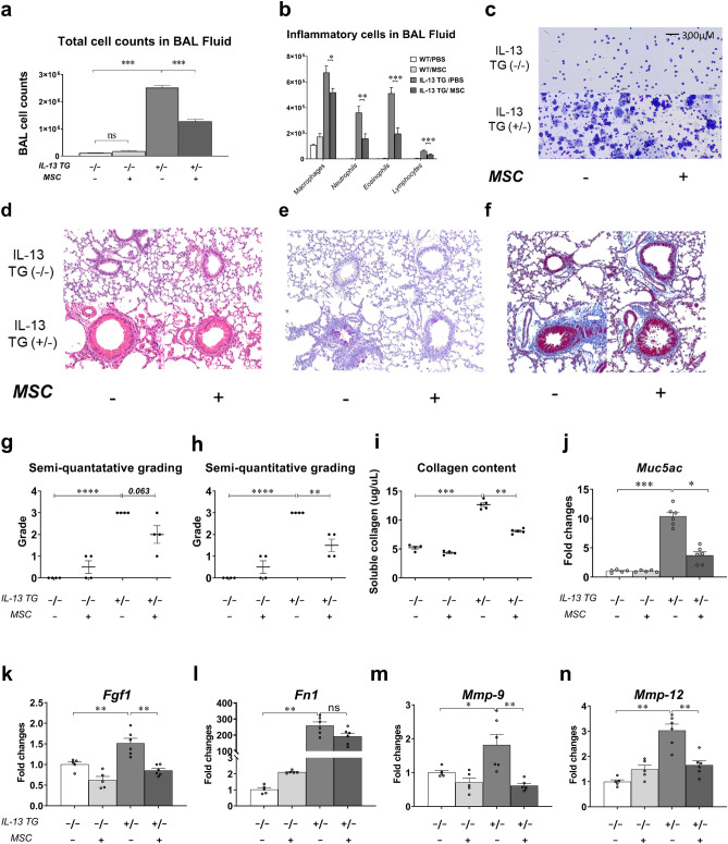
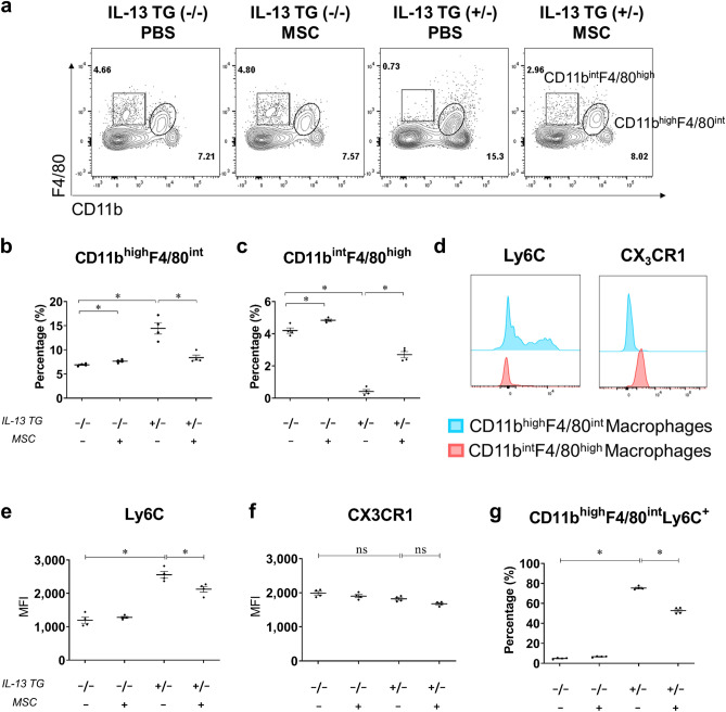
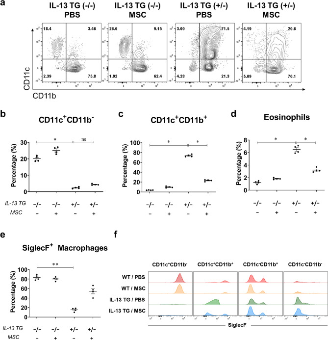
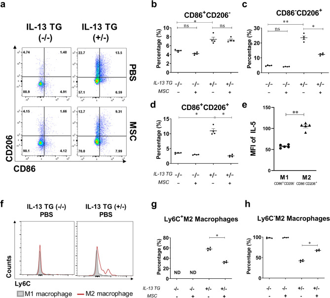
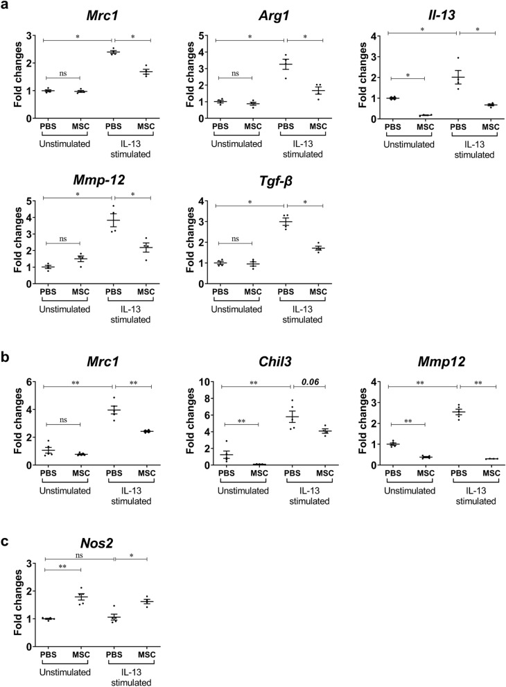
Despite numerous previous studies, the full action mechanism of the pathogenesis of asthma remains undiscovered, and the need for further investigation is increasing in order to identify more effective target molecules. Recent attempts to develop more efficacious treatments for asthma have incorporated mesenchymal stem cell (MSC)-based cell therapies. This study aimed to evaluate the anti-asthmatic effects of MSCs primed with Liproxstatin-1, a potent ferroptosis inhibitor. In addition, we sought to examine the changes within macrophage populations and their characteristics in asthmatic conditions. Seven-week-old transgenic mice, constitutively overexpressing lung-specific interleukin (IL)-13, were used to simulate chronic asthma. Human umbilical cord-derived MSCs (hUC-MSCs) primed with Liproxstatin-1 were intratracheally administered four days prior to sampling. IL-13 transgenic mice demonstrated phenotypes of chronic asthma, including severe inflammation, goblet cell hyperplasia, and subepithelial fibrosis. Ly6C+M2 macrophages, found within the pro-inflammatory CD11c+CD11b+ macrophages, were upregulated and showed a strong correlation with lung eosinophil counts. Liproxstatin-1-primed hUC-MSCs showed enhanced ability to downregulate the activation of T helper type 2 cells compared to naïve MSCs in vitro and reduced airway inflammation, particularly Ly6C+M2 macrophages population, and fibrosis in vivo. In conclusion, intratracheal administration is an effective method of MSC delivery, and macrophages hold great potential as an additional therapeutic target for asthma.
© 2022. The Author(s).
Conflict of interest statement
The authors declare no competing interests.
Figures
Similar articles
-
Mesenchymal Stem Cells Attenuate Asthmatic Inflammation and Airway Remodeling by Modulating Macrophages/Monocytes in the IL-13-Overexpressing Mouse Model.Immune Netw. 2022 Sep 27;22(5):e40. doi: 10.4110/in.2022.22.e40. eCollection 2022 Oct. Immune Netw. 2022. PMID: 36381962 Free PMC article.
-
Intravenous Mesenchymal Stem Cell Administration Modulates Monocytes/Macrophages and Ameliorates Asthmatic Airway Inflammation in a Murine Asthma Model.Mol Cells. 2022 Nov 30;45(11):833-845. doi: 10.14348/molcells.2022.0038. Epub 2022 Nov 11. Mol Cells. 2022. PMID: 36380733 Free PMC article.
-
Intratracheal administration of mesenchymal stem cells modulates lung macrophage polarization and exerts anti-asthmatic effects.Sci Rep. 2022 Jul 11;12(1):11728. doi: 10.1038/s41598-022-14846-y. Sci Rep. 2022. PMID: 35821386 Free PMC article.
-
Old Friends with Unexploited Perspectives: Current Advances in Mesenchymal Stem Cell-Based Therapies in Asthma.Stem Cell Rev Rep. 2021 Aug;17(4):1323-1342. doi: 10.1007/s12015-021-10137-7. Epub 2021 Mar 1. Stem Cell Rev Rep. 2021. PMID: 33649900 Free PMC article. Review.
-
[Mechanism of Mesenchymal Stem Cells in the Treatment of Asthma].Zhongguo Yi Xue Ke Xue Yuan Xue Bao. 2022 Oct;44(5):845-856. doi: 10.3881/j.issn.1000-503X.14960. Zhongguo Yi Xue Ke Xue Yuan Xue Bao. 2022. PMID: 36325782 Review. Chinese.
Cited by
-
Mesenchymal Stem/Stromal Cells in Asthma Therapy: Mechanisms and Strategies for Enhancement.Cell Transplant. 2023 Jan-Dec;32:9636897231180128. doi: 10.1177/09636897231180128. Cell Transplant. 2023. PMID: 37318186 Free PMC article. Review.
-
Programmed Cell Death in Asthma: Apoptosis, Autophagy, Pyroptosis, Ferroptosis, and Necroptosis.J Inflamm Res. 2023 Jul 1;16:2727-2754. doi: 10.2147/JIR.S417801. eCollection 2023. J Inflamm Res. 2023. PMID: 37415620 Free PMC article. Review.
-
CX3CR1 regulates the development of renal interstitial fibrosis through macrophage polarization.Zhong Nan Da Xue Xue Bao Yi Xue Ban. 2023 Jul 28;48(7):957-966. doi: 10.11817/j.issn.1672-7347.2023.220601. Zhong Nan Da Xue Xue Bao Yi Xue Ban. 2023. PMID: 37724398 Free PMC article. Chinese, English.
-
Paraoxonase-1 Is a Pivotal Regulator Responsible for Suppressing Allergic Airway Inflammation Through Adipose Stem Cell-Derived Extracellular Vesicles.Int J Mol Sci. 2024 Nov 27;25(23):12756. doi: 10.3390/ijms252312756. Int J Mol Sci. 2024. PMID: 39684469 Free PMC article.
-
Extracellular Vesicles in Asthma: Intercellular Cross-Talk in TH2 Inflammation.Cells. 2025 Apr 3;14(7):542. doi: 10.3390/cells14070542. Cells. 2025. PMID: 40214495 Free PMC article. Review.
References
Publication types
MeSH terms
Substances
LinkOut - more resources
Full Text Sources
Medical
Molecular Biology Databases
Research Materials
