. 2022 Dec;29(12):1854-1865.
doi: 10.1038/s41417-022-00484-z. Epub 2022 Jun 14.

Regional infusion of a class C TLR9 agonist enhances liver tumor microenvironment reprogramming and MDSC reduction to improve responsiveness to systemic checkpoint inhibition

Affiliations

Regional infusion of a class C TLR9 agonist enhances liver tumor microenvironment reprogramming and MDSC reduction to improve responsiveness to systemic checkpoint inhibition

Chandra C Ghosh et al. Cancer Gene Ther. 2022 Dec.

Abstract

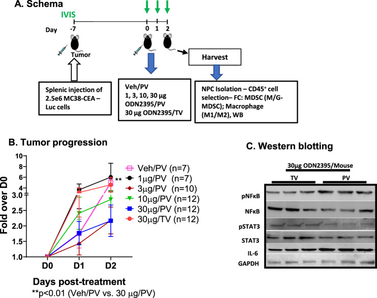
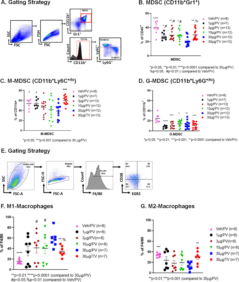
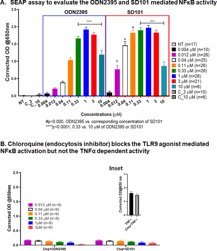
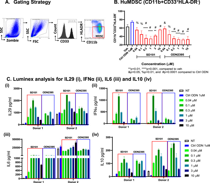
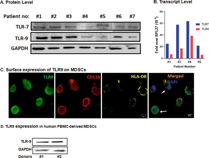
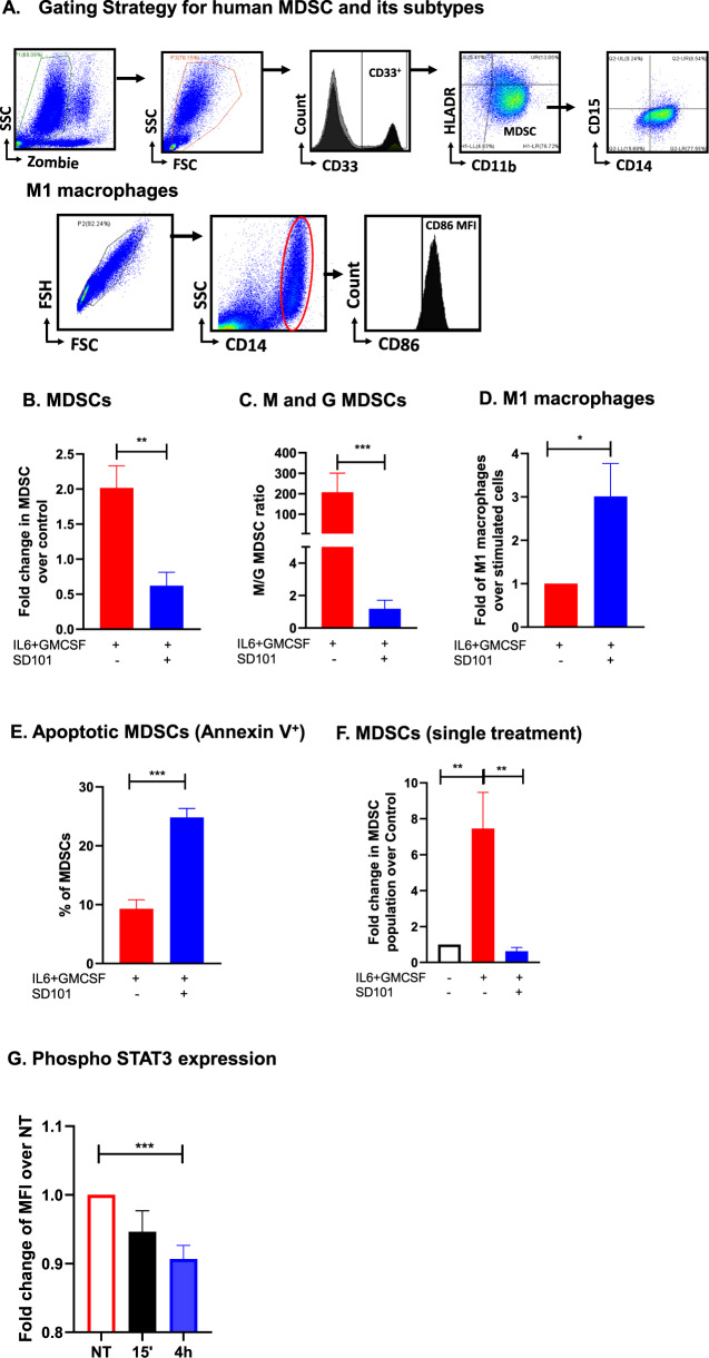
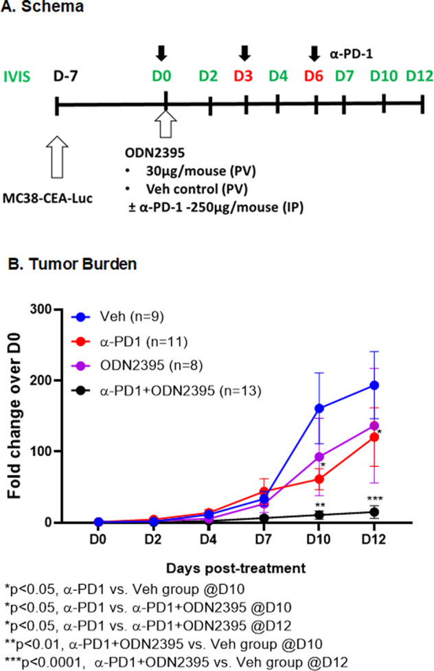
Myeloid-derived suppressor cells (MDSCs) expand in response to malignancy and suppress responsiveness to immunotherapy, including checkpoint inhibitors (CPIs). Within the liver, MDSCs have unique immunosuppressive features. While TLR9 agonists have shown promising activities in enhancing CPI responsiveness in superficial tumors amenable to direct needle injection, clinical success for liver tumors with TLR9 agonists has been limited by delivery challenges. Here, we report that regional intravascular infusion of ODN2395 into mice with liver metastasis (LM) partially eliminated liver MDSCs and reprogrammed residual MDSC. TLR9 agonist regional infusion also induced an increase in the M1/M2 macrophage ratio. Enhanced TLR9 signaling was demonstrated by an increased activation of in NFκB (pP65) and production of IL6 compared with systemic infusion. Further, PBMC-derived human MDSCs express TLR9, and treatment with class C TLR9 agonists (ODN2395 and SD101) reduced the expansion of MDSC population. TLR9 stimulation induced MDSC apoptosis and increased the M1/M2 macrophage ratio. Regional TLR9 agonist infusion along with systemic anti-PD-1 therapy improved control of LM. With effective delivery, TLR9 agonists have the potential to favorably reprogram the liver TME through reduction of MDSCs and favorable macrophage polarization, which may improve responsiveness to systemic CPI therapy.


Conflict of interest statement

SCK was a paid member of the scientific advisory board and received grant funding from TriSalus™ Life Sciences, Inc during the conduct of the research and initial drafting, and currently is employed by the company. BFC, CCG, JL, and PG are currently employed by TriSalus™ Life Sciences, Inc. The other authors have declared that no conflict of interest exists.

Figures

Fig. 1
Fig. 1. ODN2395 administered via PV is more effective in inhibiting tumor progression.
A. Schema: Schematic representation of the timeline of LM generation and the treatment protocol. Eight- to twelve-week-old C57/BL6 mice were challenged using intra-splenic injection model with 2.5 × 106 MC38-CEA-Luc cells for 7 days (D-7). Bioluminescence value was determined by IVIS on D0, D1, D2, and mice were randomized accordingly and treated with 1, 3, 10, or 30 µg/mouse ODN2395 via portal vein (PV) and 30 µg/mouse ODN2395 via tail vein (TV). PBS served as the vehicle (Veh) control and administered via PV. On D2 post-treatment with ODN2395 or Veh, mice were sacrificed, and liver was harvested to isolate CD45+ cells. Isolated CD45+ NPCs were evaluated for MDSCs and macrophages (M1 and M2). B Tumor progression was monitored by IVIS imaging on the day of treatment (D0), D1, and D2 post-treatment. Fold change of the tumor burden was calculated based on D0 baseline bioluminescence. Multiple t test was performed to determine the significant difference. C Harvested LM tissues (whole lysates) from n = 6 mice/group (representative of n = 3 shown) were evaluated for pNFκB (p65S536), pSTAT3Y705, total NFκB, STAT3, and IL6 by western blotting. GAPDH was used as a housekeeping protein control.
Fig. 2
Fig. 2. ODN2395 administered via PV reprograms the myeloid cell phenotype in the LM.
A As described in Fig. 1A, mice were sacrificed after 2 days post-treatment. CD45+ cells were isolated from non-parenchymal cells (NPCs). Gating strategy to analyze CD45+ cells isolated from the LM. B MDSC cell population (CD11b+Gr1+), C monocytic MDSCs (M-MDSC; CD11b+Ly6C+/hiLy6G/lo) and D granulocytic MDSCs (G-MDSC; CD11b+Ly6C-/loLy6G+/hi) were measured. E Gating strategy of the phenotypic analysis of CD45+ derived macrophages that were isolated from the LM. F M1-macrophage cell population (F4/80+CD38+Egr2-) and G M2 macrophage cell population (F4/80+CD38-Egr2+) were determined. Each animal data is represented by a scattered plot and presented as mean ± SEM from at least three different experiments. Students’ t-test was performed for group-wise comparison and are described in each graph.
Fig. 3
Fig. 3. Determination of TLR9-dependent activity of TLR9 agonists.
A In this reporter-based assay, TLR9-expressing HEK293-Blue cells were treated with ODN2395 and SD101 at increasing doses (0.004–10 µM) for 21 h. As a negative control, no-treatment (NT) and sequence control ODN5328 at 3 (C_3) and 10 (C_10) µM were used. The secreted embryonic alkaline phosphatase (SEAP) was determined by measuring the absorbance at 650 nm after addition of substrate. B Cells were pretreated with chloroquine (Chq, 1 µg/ml) for 45 min before the addition of ODN2395 or SD101 at increasing concentrations (0.012–3 µM) for 21 h, and absorbance was measured at 650 nm. All the experiments were performed at least three times with 2–3 replicates, and mean ± SEM was plotted in the graph.
Fig. 4
Fig. 4. TLR9 agonist-treated huPBMC reduced MDSC population and increases IFNα- and NFκB-regulated genes.
A Gating strategy for phenotypic analysis of MDSCs; isolated human PBMCs were treated with increasing concentrations (0.04–10 µM) of SD101, ODN2395 along with ctrl ODN5328 (1 µM) for 48 h. B MDSC population was quantified followed by FC analysis. Four donors with three replicates were used, and data represented as mean ± SEM (n = 12). C Supernatants of Donor 1 and 2 were collected analyzed for (i) IL29, (ii) IFNα, (iii) IL6, (iv) IL10 by using Luminex. Cells treated with SD101 and ODN2395 are represented as red and blue boxes, respectively. Data from two representative donors with two replicates are presented here. For Donor 1 and 2 supernatants from 10 µM ODN2395 treated samples were unavailable for luminex potential analysis.
Fig. 5
Fig. 5. Expression of TLR9 in LM biospecimens and MDSCs.
A Protein lysates obtained from LM patient biospecimens were evaluated for TLR7 and TLR9 by western blotting. GAPDH was used as housekeeping protein control. WB was performed on two different runs (#1 to #5 on one run and #6 and #7 were performed in a different run) B Total RNA was isolated from the same biospecimen and expression of TLR9 was quantified by qRT-PCR. RPL-27 gene was used as housekeeping control. Representative data of 4 of 7 biospecimens is shown here due to sample unavailability). C IL6 (20 ng/ml) + GMCSF (20 ng/ml) stimulated PBMCs grown in chamber slides were fixed and stained with TLR9 (green), CD11b (red) and HLA-DR (yellow) antibodies and DAPI (blue) used for nuclear staining. IF images demonstrate surface expression of TLR9 in CD11b+HLA-DR cells. Representative of three different experiment using PBMCs from three different donors. D WB of IL6 (20 ng/ml) + GMCSF (20 ng/ml) treated PBMCs showing expression of TLR9. GAPDH was used as control. (Representative of two out of four donors).
Fig. 6
Fig. 6. SD101 inhibits the generation of huMDSCs from PBMCs.
A Gating strategy to identify huMDSCs, its subtypes M- and G-MDSCs and M1 macrophages. PBMCs were treated with IL6 (20 ng/ml) + GMCSF (20 ng/ml) for 7 days in the presence or absence of 0.3 µM SD101. B Cells were treated with SD101 on D0, D2, and D7 and percentage of MDSCs (CD11b+CD33+HLA-DRlo/−) was measured. C Ratio of M-/G-MDSCs was quantified; M-MDSCs: CD11b+CD14+CD15-HLA-DRlo/−, G-MDSCs:CD11b+CD14-CD15+HLA-DRlo/−. D Macrophage population was quantified: CD14+CD86+. E Annexin positive MDSCs were quantified. F PBMCs were treated once on D0 with SD101 (0.3 µM) for 48 h and MDSC was quantified. G PBMCs were stimulated with IL6 + GMCSF and treated with SD101 for 15 min and 4 h. FC analysis was performed to quantify pSTAT3 MFI in the MDSC gated cells and reported as fold change in the MFI of pSTAT3 positive cells. All the experiments were performed at least three times, and mean ± SEM was plotted in the graph.
Fig. 7
Fig. 7. Locoregional TLR9 stimulation with systemic CPI effectively inhibits tumor progression.
A Schema illustrates the steps to develop LM and the treatment regimen. Eight- to twelve-week-old male C57/BL6 mice were challenged using intra-splenic injection model with MC38-CEA-Luc cells for 1 week. Bioluminescence was measured by IVIS and mice were randomized on D0 prior to treatment with 30 µg/mouse ODN2395 via PV with or without 250 µg/mouse of anti-PD1 antibody via IP on D0, D3, and D6. PBS-treated mice via PV were used as control. B Tumor growth was monitored by IVIS imaging on D2, D4, D7, D10, and D12. Fold change of the tumor burden was calculated based on D0 baseline bioluminescence. Data was analyzed by multiple t test. Discovery determined using the two-stage linear step-up procedure of Benjamini, Krieger and Yekutieli, with Q = 1%.

References

    1. Yu J, Green MD, Li S, Sun Y, Journey SN, Choi JE, et al. Liver metastasis restrains immunotherapy efficacy via macrophage-mediated T cell elimination. Nat Med. 2021;27:152–64. doi: 10.1038/s41591-020-1131-x. - DOI - PMC - PubMed
    1. Pelster MS, Gruschkus SK, Bassett R, Gombos DS, Shephard M, Posada L, et al. Nivolumab and ipilimumab in metastatic uveal melanoma: results from a single-arm phase II study. J Clin Oncol. 2021;39:599–607. doi: 10.1200/JCO.20.00605. - DOI - PMC - PubMed
    1. Blum ES, Yang J, Komatsubara KM, Carvajal RD. Clinical management of uveal and conjunctival melanoma. Oncol (Williston Park) 2016;30:29–32. - PubMed
    1. Chattopadhyay C, Kim DW, Gombos DS, Oba J, Qin Y, Williams MD, et al. Uveal melanoma: From diagnosis to treatment and the science in between. Cancer. 2016;122:2299–312. doi: 10.1002/cncr.29727. - DOI - PMC - PubMed
    1. Sussman TA, Funchain P, Singh A. Clinical trials in metastatic uveal melanoma: current status. Ocul Oncol Pathol. 2020;6:381–7. doi: 10.1159/000508383. - DOI - PMC - PubMed