. 2022 Nov;40(11):1680-1689.
doi: 10.1038/s41587-022-01347-6. Epub 2022 Jun 13.

Rapid, scalable assessment of SARS-CoV-2 cellular immunity by whole-blood PCR

Affiliations

Rapid, scalable assessment of SARS-CoV-2 cellular immunity by whole-blood PCR

Megan Schwarz et al. Nat Biotechnol. 2022 Nov.

Abstract

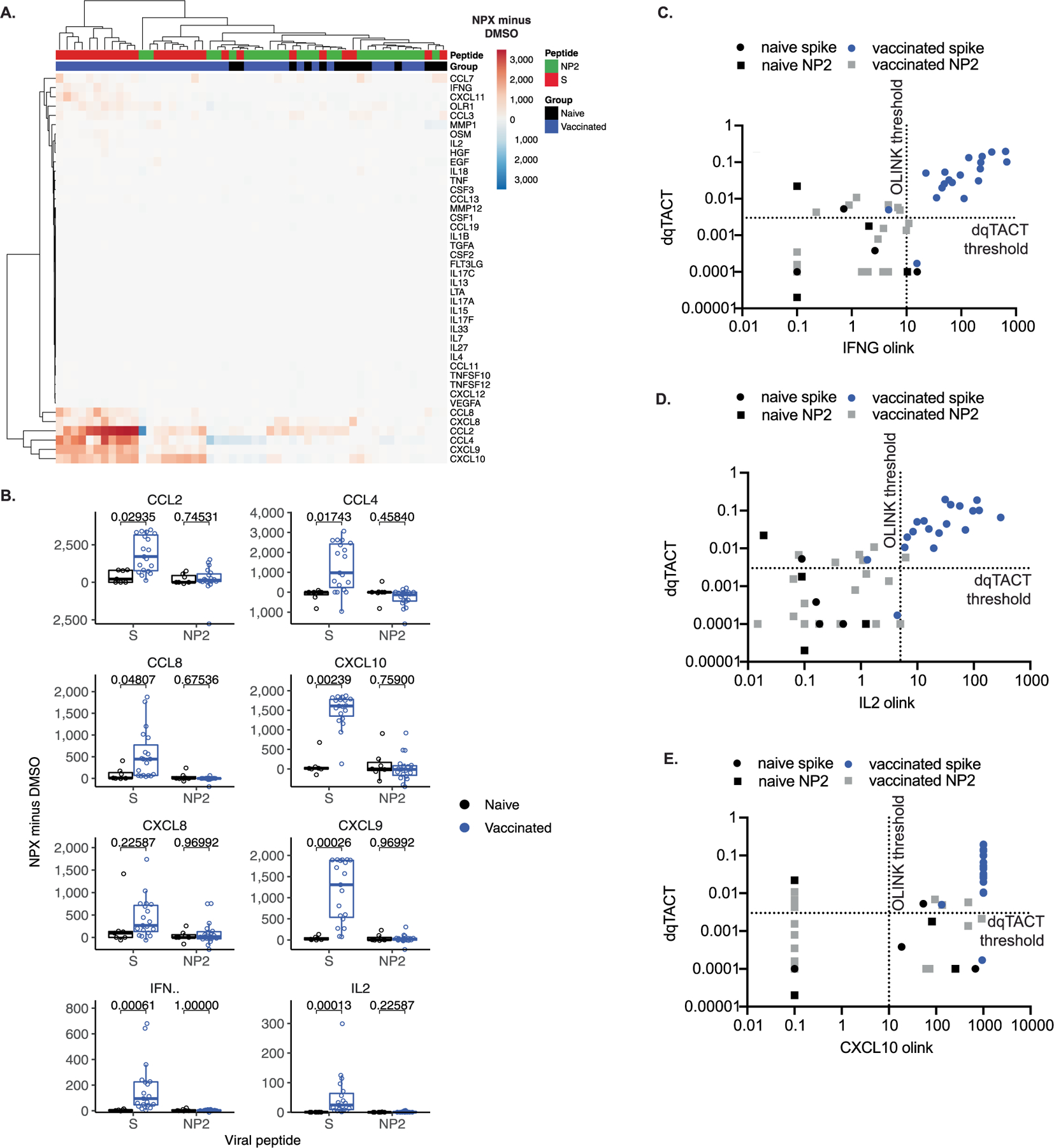
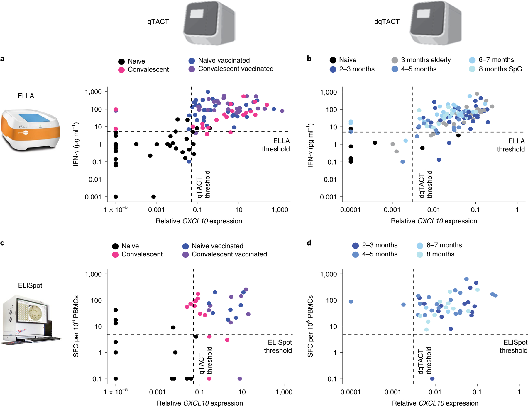
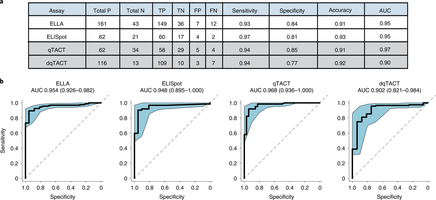
Fast, high-throughput methods for measuring the level and duration of protective immune responses to SARS-CoV-2 are needed to anticipate the risk of breakthrough infections. Here we report the development of two quantitative PCR assays for SARS-CoV-2-specific T cell activation. The assays are rapid, internally normalized and probe-based: qTACT requires RNA extraction and dqTACT avoids sample preparation steps. Both assays rely on the quantification of CXCL10 messenger RNA, a chemokine whose expression is strongly correlated with activation of antigen-specific T cells. On restimulation of whole-blood cells with SARS-CoV-2 viral antigens, viral-specific T cells secrete IFN-γ, which stimulates monocytes to produce CXCL10. CXCL10 mRNA can thus serve as a proxy to quantify cellular immunity. Our assays may allow large-scale monitoring of the magnitude and duration of functional T cell immunity to SARS-CoV-2, thus helping to prioritize revaccination strategies in vulnerable populations.


Conflict of interest statement

Competing interests

A.B., A.T. and N.L.B. declare the filling of a patent application relating to the use of peptide pools in whole blood for detection of SARS-CoV-2 T cells (pending). E.G., J.O., M.S., D.T. and D.L.O. declare the filling of a patent application relating to the qTACT and dqTACT assays (pending). S.G. reports consultancy and/or advisory roles for Merck and OncoMed, and research funding from Bristol-Myers Squibb, Celgene, Genentech, Immune Design, Janssen R&D, Pfizer, Regeneron and Takeda. G.N., I.D.N. and L.C. are employees of Hyris Ltd, manufacturer of the bCUBE machine described in this article. C.L., G.S. and M.F. are employees of Synlab Italy. The other authors declare no competing interests.

Figures

Extended Data Fig. 1 |
Extended Data Fig. 1 |. CXCL10 mRNA levels can correlate with IFNG mRNA levels and can be reliably used as a proxy for T cell activation.
A. Normalized counts of candidate genes selected for downstream validation based on differential expression versus DMSO. Comparisons show significance calculated using DESeq2 (two-sided) and corrected using the Benjamini-Hochberg method. Naive, N = 11; convalescent N = 8; vaccinated, N = 16 biologically independent samples. B. Correlation between candidate gene and IFNG expression (normalized gene counts). Treatment and group is indicated by color/symbol (see legend). Correlation R and p-values were calculated using Spearman’s index. Grey bands represent the 95% confidence interval of the linear model. C. Concordance between ELLA and TACTseq NGS assays. For 24 subjects, IFN-γ protein secretion was quantified by ELLA (y-axis) and CXCL10 mRNA by Illumina NGS sequencing TACTseq (x-axis). D. Validation of candidate genes in naive (black) and convalescent (pink) subjects stimulated with NP2 or spike SpG peptide pools (qTACT assay). Naive, N = 13; convalescent, N = 16 biologically independent samples. P-values were calculated using a two-sided Wilcoxon rank sum method E. Average normalized variance in relative CXCL10 expression minus DMSO across replicate qTACT runs on the same samples. For A and D, the box bounds represent the first quartile (bottom), median (center) and the third quartile (top). The whiskers represent the range of samples up to 1.5 times the interquartile range. Beyond this point, the samples are shown as outliers.
Extended Data Fig. 2 |
Extended Data Fig. 2 |. Gating strategies for flow cytometry experiments.
a. Gating strategy for main Fig. 2 panel B. b. Gating strategy for main Fig. 2 panel D.
Extended Data Fig. 3 |
Extended Data Fig. 3 |. Comparing TACTseq assay to other T cell assays and dqTACT on different PCR machines.
A. Concordance between ELLA and TACTseq assays. For 24 subjects, IFN-γ secretion was quantified by ELLA (y-axis) and CXCL10 mRNA by TACTseq (x-axis). The quantification shown is with the DMSO control subtracted from the spike peptide stimulated sample. Each dot represents a unique subject color coded based on their COVID-19 and vaccination statuses. The dashed line represents thresholds for each assay. B. Concordance between ELISpot and TACTseq assays. For 24 subjects, IFN-γ producing cells were quantified by ELISpot (y-axis) and CXCL10 mRNA by TACTseq (x-axis). The quantification shown is with the DMSO control subtracted from the spike peptide stimulated sample. Each dot represents a unique subject color coded based on their COVID-19 and vaccination statuses. The dashed line represents thresholds for each assay. C. Concordance between ELISpot and ELLA assays. For 85 samples, IFN-γ protein secretion was quantified by ELLA (y-axis) and IFN-γ producing cells were quantified by ELISpot (y-axis). The quantification shown is with the DMSO control subtracted from the spike peptide stimulated sample. Each dot represents a unique subject color coded based on their COVID-19 and vaccination statuses. The dashed line represents thresholds for each assay. D. Nonparametric Spearman’s correlation between dqTACT samples run on Bio-Rad CFX384 (y-axis) and Hyris bCUBE 2.0 (x-axis). Bands represent 95% confidence interval. E. Nonparametric Spearman’s correlation between dqTACT samples run on Bio-Rad CFX96 (y-axis) and Applied Biosystems AB 7500 (x-axis). Bands represent 95% confidence interval.
Extended Data Fig. 4 |
Extended Data Fig. 4 |. Comparing dqTACT and Olink assays.
A. Heatmap displaying normalized protein expression (NPX) values minus DMSO across 42 peptides measured by Olink. B. Boxplot displaying normalized protein expression (NPX) values minus DMSO. Comparisons show significance for the Wilcoxon Rank Sum two-sided test, corrected using the Benjamini-Hochberg method. Naive, N = 7; vaccinated, N = 19 biologically independent samples. The box bounds represent the first quartile (bottom), median (center), and the third quartile (top). The whiskers represent the range of samples up to 1.5 times the interquartile range. Beyond this point, samples are shown as outliers. C-E. Concordance between Olink and dqTACT assays. For 23 samples, IFN-γ (C), IL2 (D), and CXCL10 (E) protein secretion was quantified by O-link (x-axis) and CXCL10 mRNA by qTACT (y-axis). The quantification shown is with the DMSO control subtracted from the spike peptide stimulated sample. Each dot represents a unique subject color coded based on their COVID-19 and vaccination statuses. The dashed line represents thresholds for each assay.
Extended Data Fig. 5 |
Extended Data Fig. 5 |. Using the dqTACT assay for a large clinical trial cohort (CombiVacS).
A. Concordance between ELLA and qTACT assays for IFN-γ/IFNG. For n = 91 subjects, IFN-γ secretion was quantified by ELLA (x-axis) and IFNG mRNA by qTACT (y-axis). Colors on their COVID-19 and vaccination statuses (see legend). The dashed lines represent thresholds for each assay. B-C. Quantification CXCL10 mRNA using dqTACT (B) or IFN-γ protein section using ELLA (C) in subjects enrolled in the CombiVacS trial. All subjects received a first dose of ChAdOx1s from AstraZeneca (Dose 1 (AZ)). The patients were then divided into two groups and received either a second placebo dose (Dose 1 (AZ) + Dose 2 (placebo)) or a second dose of BNT162b2 from Pfizer (Dose 1 (AZ) + Dose 2 (Pfizer)). The dqTACT assay was completed as shown in Fig. 1A (bottom). Comparisons show significance for the Wilcoxon rank sum two-sided test. For (B), naive, N = 13; dose 1 (AZ), N = 142, dose 1 (AZ) + dose 2 (placebo), N = 49; dose 1 (AZ) + dose 2 (Pfizer), N = 92 biologically independent samples. For (C), naive, N = 43; dose 1 (AZ), N = 155, dose 1 (AZ) + dose 2 (placebo), N = 52; dose 1 (AZ) + dose 2 (Pfizer), N = 99 biologically independent samples.
Fig. 1 |
Fig. 1 |. CXCL10 mRNA levels can be measured as a proxy for T cell activation.
a, Schematic of workflow for the T cell activation assays. All assays begin with whole-blood collection followed by overnight stimulation with DMSO, nucleocapsid (NP) or SpG peptide pools. Next, supernatants are collected for ELLA or Olink; RNA is extracted and used for probe-based qPCR (qTACT) or next-generation sequencing (TACTseq) or whole blood is diluted and used directly for qPCR (dqTACT). Figure created with Biorender.com. b, CXCL10 is upregulated in response to spike peptide pool activation of whole blood. Volcano plot displaying differentially expressed genes stimulated by the spike peptide pool versus DMSO, grouped by the participant’s COVID-19 or vaccination status, displayed as red (upregulated) or blue (downregulated). Significantly differentially expressed genes were defined as having an adjusted P value <0.05 and |log2FC| > 1. P values were calculated using DESeq2 (two-sided) and adjusted using the Benjamini–Hochberg method. c, CXCL10 and IFNG mRNA induction correlate. Scatter plot displaying each gene’s correlation to IFNG expression across samples (x axis), and the corresponding log2FC (calculated by DESeq2) for the spike peptide pool versus DMSO, grouped by COVID-19 or vaccination status. Correlation calculated using Spearman’s index. d, Venn diagram displaying overlap in significantly upregulated genes in convalescent and vaccinated participants for spike peptide-stimulated samples compared to DMSO control samples. Significantly upregulated genes were defined as having P < 0.05 and log2FC > 1. e, GSEA plot displaying significantly positive enrichment for IFN-γ response genes among the upregulated genes in spike peptide simulate samples compared to DMSO control samples in COVID-19 convalescent and vaccinated cohorts.
Fig. 2 |
Fig. 2 |. CXCL10 is upregulated by monocytes in response to IFN-γ released by antigen-specific T cells.
a, Schematic of proposed mechanism of CXCL10 transcript upregulation. On spike stimulation of whole blood, antigen presenting cells (APCs) present spike peptides to antigen-specific T cells that subsequently release IFN-γ. IFN-γ release can be quantified by ELLA, ELISpot or flow cytometry. Next, IFN-γ stimulates monocytes, which, in turn, upregulate CXCL10 mRNA, which can be detected by the qTACT/dqTACT assays. Figure created with Biorender.com. b, Only monocytes upregulate CXCL10 in response to IFN-γ and TNF-α. Whole blood was stimulated with DMSO (Neg) or IFN-γ + TNF-α in the presence of brefeldin/Monensin (BFA/Mon). CXCL10/IP-10 positive T cells (T), B cells (B), natural killer cells (NK), natural killer T cells (NK-T), monocytes (Mono) and neutrophils (Neutro) were quantified using flow cytometry. P values were calculated using a two-sided Wilcoxon matched-pairs signed rank test. n = 4 biologically independent samples. c, Both monocytes and neutrophils release CXCL10/IP-10 on SpG stimulation. Whole blood was stimulated with DMSO (Neg) or a spike peptide pool (SpG) in the absence (left panel) or presence (right panel) of BFA/Mon. CXCL10 positive T, B, NK, NK-T, Mono and Neutro were quantified using flow cytometry. P values were calculated using a two-sided Wilcoxon matched-pairs signed rank test. n = 4 biologically independent samples. d, Monocytes, and not neutrophils, upregulate CXCL10/IP-10 in response to SpG. Whole blood was simulated with DMSO (Neg) or a spike peptide pool (SpG) overnight with BFA/Mon added for the last 4 h. CXCL10 positive Monocytes and Neutrophils were quantified using flow cytometry. P values were calculated using a two-sided Wilcoxon matched-pairs signed rank test (P = 0.015625). n = 7 biologically independent samples. e, CXCL10 mRNA is upregulated in monocytes on stimulation with spike peptides. Monocytes and neutrophils were sorted from whole blood after overnight stimulation with spike peptides or DMSO control. RNA was extracted from the cells and the relative CXCL10 mRNA expression was determined using the qTACT assay. P values were calculated using a two-sided Wilcoxon matched-pairs signed rank test (P = 0.03125). n = 7 biologically independent samples. For be, the box bounds represent the first quartile (bottom), median (center) and the third quartile (top). The whiskers represent the range of samples up to 1.5 times the interquartile range. Beyond this point, samples are shown as outliers.
Fig. 3 |
Fig. 3 |. TACT assays are concordant with gold standard ELLA and ELISpot assays.
ad, Concordance between assays to quantify cellular immunity. The quantification shown is with the DMSO control subtracted from the spike peptide-stimulated sample. Each dot represents a unique participant color coded based on their COVID-19 and vaccination statuses (legend). The dashed line represents thresholds for each assay. a, Concordance between ELLA and qTACT assays. For 117 participants, IFN-γ protein secretion was quantified by ELLA (y axis) and CXCL10 mRNA by qTACT (x axis). b, Concordance between ELLA and dqTACT assays. For 133 participants, IFN-γ protein secretion was quantified by ELLA (y axis) and CXCL10 mRNA by dqTACT (x axis). c, Concordance between ELISpot and qTACT assays. For 50 participants, IFN-γ producing cells were quantified by ELISpot (y axis) and CXCL10 mRNA by qTACT (x axis). d, Concordance between ELISpot and dqTACT assays. For 46 participants, IFN-γ producing cells were quantified by ELISpot (y axis) and CXCL10 mRNA by dqTACT (x axis).
Fig. 4 |
Fig. 4 |. Analytical validation and comparison of available T cell assays.
a, Comparison of the various assays used to determine T cell response to spike peptides. Total P/N = total positives/negatives (that is, above or below threshold, respectively). TP/TN = true positives/negatives (that is, correctly above or below threshold, respectively, according to the participant’s COVID-19/vaccination status). FP/FN = false positives/negatives. Sensitivity = true positives/(true positives + false negatives). Specificity = true negatives/(true negatives + false positives). Diagnostic accuracy = (true positives + true negatives)/all samples). b, ROC curves, AUC values and associated 95% confidence intervals for each assay.
Fig. 5 |
Fig. 5 |. Using qTACT to monitor cellular immunity in vaccinated participants.
a, Quantification of CXCL10 mRNA (qTACT) prevaccination and at 10 and 20 days after the first and second dose of an mRNA-based vaccine. Time points are indicated on the x axis and relative CXCL10 expression (minus DMSO control) on the y axis. The dashed line represents the qTACT threshold (0.05). The number of participants for each time point is indicated above the box plots along with the percentage of participants who fall above the threshold. Colors represent the participant’s COVID-19/vaccination status (legend). b, Quantification of IFNG mRNA (qTACT) prevaccination and at 10 and 20 days after the first and second dose of an mRNA-based vaccine. Time points are indicated on the x axis and relative IFNG expression (minus DMSO control) on the y axis. The number of participants for each time point is indicated above the box plots. Colors represent the participant’s COVID-19/vaccination status (legend). c, Quantification of IFN-γ protein secretion (ELLA) prevaccination and at 10 and 20 days post the first and second dose of an mRNA-based vaccine. Time points are indicated on the x axis and IFN-γ protein secretion (minus DMSO control) on the y axis. The dashed line represents the ELLA threshold (5). The number of participants for each time point is indicated above the box plots along with the percentage of participants who fall above the threshold. Colors represent the participants’ COVID-19/vaccination statuses (legend). d, Quantification of IFN-γ producing cells (ELISpot) prevaccination and at 10 and 20 days after the first and second dose of an mRNA-based vaccine. Time points are indicated on the x axis and number of IFN-γ producing cells (minus DMSO control) on the y axis. The dashed line represents the ELISpot threshold (5). The number of participants for each time point is indicated above the box plots along with the percentage of participants who fall above the threshold. Colors represent the participants’ COVID-19/vaccination statuses (legend). For a–d, the box bounds represent the first quartile (bottom), median (center) and the third quartile (top). The whiskers represent the range of samples up to 1.5 times the interquartile range. Beyond this point, samples are shown as outliers.
Fig. 6 |
Fig. 6 |. Using dqTACT to monitor the persistence of cellular immunity and cross reactivity with spike epitopes from VOC in vaccinated participants.
ac, Detection of CXCL10 mRNA (dqTACT (a)), IFN-γ protein secretion (ELLA (b)) or IFN-γ producing cells (ELISpot (c)) in vaccinated participants over time. Time points (postvaccination) are indicated on the x axis and relative CXCL10 expression (a), IFN-γ protein secretion (b) or IFN-γ producing cells (c) on the y axis (all values minus DMSO). The dashed lines represent the dqTACT (0.003), ELLA (5) or ELISpot (5) thresholds. d, Schematic to show how T cell responses against the VOC can be evaluated using the delta variant as an example. Orange regions refer to amino acid mutations present in the delta variant compared to the wildtype (WT) SARS-CoV-2 strain. Pool HS (hotspot) contains peptides covering the nonconserved Spike-Wuhan regions affected by mutations present in the delta variant (24 peptides). Pool delta MT contains peptides from Pool HS with the amino acid mutations present in the Spike protein of the delta variant. eg, Quantification of CXCL10 mRNA (dqTACT (e)), IFN-γ protein secretion (ELLA (f)) or IFN-γ producing cells (ELISpot (g)) in vaccinated participants stimulated with spike peptides covering the hotspot wildtype (HS WT) or delta variant region. The peptide pool used for stimulation, either the wildtype (HS) or delta (delt) sequence, is indicated on the x axis and relative CXCL10 expression (e), IFN-γ protein secretion (f) or IFN-γ producing cells (g) on the y axis (all values minus DMSO). The dashed lines represent the dqTACT (0.003), ELLA (5) or ELISpot (5) thresholds. P values were calculated using a two-sided Wilcoxon rank sum test. h,i, Quantification of relative CXCL10 mRNA expression using dqTACT (h) or IFN-γ protein section using ELLA (i) in an elderly cohort. For a–c and e–i, the box bounds represent the first quartile (bottom), median (center) and the third quartile (top). The whiskers represent the range of samples up to 1.5 times the interquartile range. Beyond this point, the samples are shown as outliers. The number of participants for each time point is indicated above the box plots along with the percentage of participants who fall above the threshold.

References

    1. Bhatraju PK et al. COVID-19 in critically ill patients in the Seattle Region—case series. N. Engl. J. Med 382, 2012–2022 (2020). - PMC - PubMed
    1. Wu C et al. Risk factors associated with acute respiratory distress syndrome and death in patients with coronavirus disease 2019 pneumonia in Wuhan, China. JAMA Intern. Med 180, 934–943 (2020). - PMC - PubMed
    1. Dagan N et al. BNT162b2 mRNA COVID-19 vaccine in a nationwide mass vaccination setting. N. Engl. J. Med 384, 1412–1423 (2021). - PMC - PubMed
    1. McMahan K et al. Correlates of protection against SARS-CoV-2 in rhesus macaques. Nature 590, 630–634 (2021). - PMC - PubMed
    1. Wajnberg A et al. Robust neutralizing antibodies to SARS-CoV-2 infection persist for months. Science 370, 1227–1230 (2020). - PMC - PubMed

Publication types