Review
. 2022 Jun 13;38(1):16.
doi: 10.1186/s42826-022-00126-3.

Real-time observation of neutrophil extracellular trap formation in the inflamed mouse brain via two-photon intravital imaging

Affiliations
Review

Real-time observation of neutrophil extracellular trap formation in the inflamed mouse brain via two-photon intravital imaging

Da Jeong Byun et al. Lab Anim Res. .

Abstract

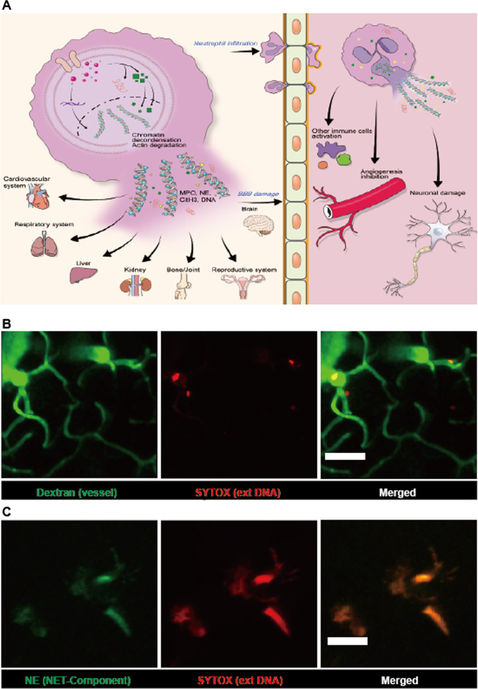
Intravital imaging via two-photon microscopy (TPM) is a useful tool for observing and delineating biological events at the cellular and molecular levels in live animals in a time-lapse manner. This imaging method provides spatiotemporal information with minimal phototoxicity while penetrating a considerable depth of intact organs in live animals. Although various organs can be visualized using intravital imaging, in the field of neuroscience, the brain is the main organ whose cell-to-cell interactions are imaged using this technique. Intravital imaging of brain disease in mouse models acts as an abundant source of novel findings for studying cerebral etiology. Neutrophil infiltration is a well-known hallmark of inflammation; in particular, the crucial impact of neutrophils on the inflamed brain has frequently been reported in literature. Neutrophil extracellular traps (NETs) have drawn attention as an intriguing feature over the last couple of decades, opening a new era of research on their underlying mechanisms and biological effects. However, the actual role of NETs in the body is still controversial and is in parallel with a poor understanding of NETs in vivo. Although several experimental methods have been used to determine NET generation in vitro, some research groups have applied intravital imaging to detect NET formation in the inflamed organs of live mice. In this review, we summarize the advantages of intravital imaging via TPM that can also be used to characterize NET formation, especially in inflamed brains triggered by systemic inflammation. To study the function and migratory pattern of neutrophils, which is critical in triggering the innate immune response in the brain, intravital imaging via TPM can provide new perspectives to understand inflammation and the resolution process.

Keywords: Brain; Intravital imaging; Neutrophil; Neutrophil extracellular trap; Two-photon microscopy.


Conflict of interest statement

The authors declare that they have no competing interests.

Figures

Fig. 1
Fig. 1
Brain intravital imaging. A Each image represents the setup of mouse brain imaging with the chamber for two-photon intravital imaging. Anesthetized mouse was put in a customized chamber to hold the skull and maintain body temperature. A craniotomy was conducted to expose the cortex surface. The exposed cortex was covered with cover glass (3–5 mm), and the metal ring was placed parallel to the brain structure (upper panels). The cranial window of the mouse brain was connected to a water-immersed lens for imaging capture. Next, the lens in the cranial window was immersed in PBS (bottom panels). B A snapshot of two-photon intravital imaging of the mouse brain is displayed. The bloodstream in this imaging was visualized with Texas Red-dextran (70 kDa, 2.5 mg/kg). Scale bar: 50 μm
Fig. 2
Fig. 2
Formation of neutrophil extracellular traps (NETs). A Schema of NET formation. In the process of NET formation, various molecular components such as ROS, PAD4, NE, MPO, and citrullinated histone are involved. Once the membrane is ruptured, the intracellular component is emitted as tangled neutrophil components. This is called a NET. It can be found in any organ and location. Thus, NETs and their accompanied components are involved in neutrophil-gated immune response in multiple organs, including the brain. The image sets represent intravital imaging of NETs in the LPS-induced inflamed mouse brain conducted via two-photon microscopy. B The brain blood vessel was stained with FITC-dextran (green, 70 kDa, 2.5 mg/kg) and SYTOX-orange (red, 5 mM). As SYTOX labels DNA strands not covered with intact membranes, it is possibly used as a NET indicator (red). Scale bar: 50 μm. C NETs are visualized using two different NET-defining markers: SYTOX-orange (red, 5 mM) and neutrophil elastase–Alexa 488 conjugated antibody (green, 0.1 mg/kg). Neutrophil elastase is used as one of the components of the NETs. It is observed as tangled with extDNA stained with SYTOX. Scale bar: 20 μm

Similar articles

Cited by

References

    1. Nwaneshiudu A, Kuschal C, Sakamoto FH, Anderson RR, Schwarzenberger K, Young RC. Introduction to confocal microscopy. J Invest Dermatol. 2012;132(12):e3. doi: 10.1038/jid.2012.429. - DOI - PubMed
    1. Yew E, Rowlands C, So PT. Application of multiphoton microscopy in dermatological studies: a mini-review. J Innov Opt Health Sci. 2014;7(5):1330010. doi: 10.1142/S1793545813300103. - DOI - PMC - PubMed
    1. Helmchen F, Denk W. Deep tissue two-photon microscopy. Nat Methods. 2005;2(12):932–40. doi: 10.1038/nmeth818. - DOI - PubMed
    1. Weigert R, Sramkova M, Parente L, Amornphimoltham P, Masedunskas A. Intravital microscopy: a novel tool to study cell biology in living animals. Histochem Cell Biol. 2010;133(5):481–91. doi: 10.1007/s00418-010-0692-z. - DOI - PMC - PubMed
    1. Konig K. Multiphoton microscopy in life sciences. J Microsc. 2000;200(Pt 2):83–104. doi: 10.1046/j.1365-2818.2000.00738.x. - DOI - PubMed

LinkOut - more resources