. 2022 Jun 14;10(1):91.
doi: 10.1186/s40168-022-01273-4.

Deletion of both Dectin-1 and Dectin-2 affects the bacterial but not fungal gut microbiota and susceptibility to colitis in mice

Affiliations

Deletion of both Dectin-1 and Dectin-2 affects the bacterial but not fungal gut microbiota and susceptibility to colitis in mice

Yazhou Wang et al. Microbiome. .

Abstract

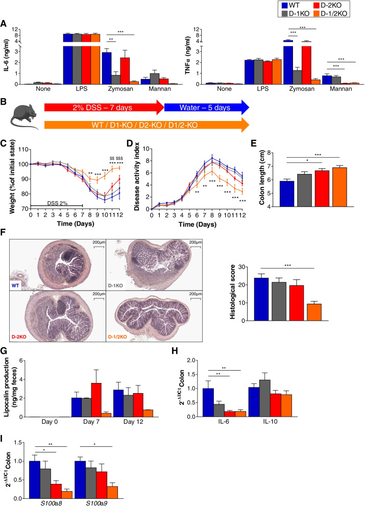
Background: Innate immunity genes have been reported to affect susceptibility to inflammatory bowel diseases (IBDs) and colitis in mice. Dectin-1, a receptor for fungal cell wall β-glucans, has been clearly implicated in gut microbiota modulation and modification of the susceptibility to gut inflammation. Here, we explored the role of Dectin-1 and Dectin-2 (another receptor for fungal cell wall molecules) deficiency in intestinal inflammation.

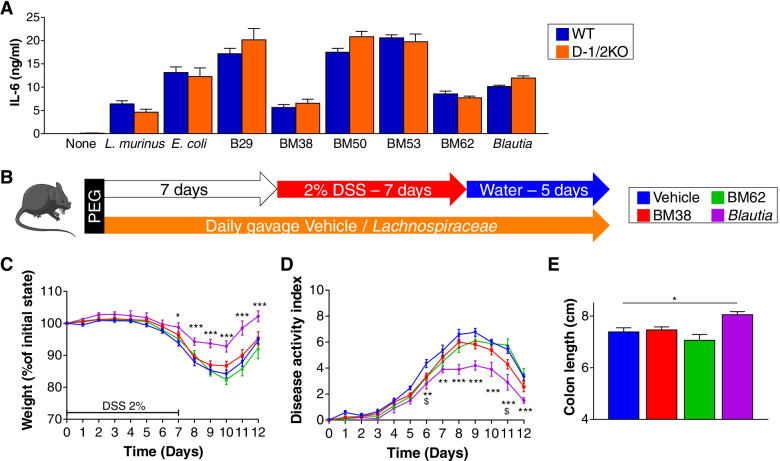
Design: Susceptibility to dextran sodium sulfate (DSS)-induced colitis was assessed in wild-type, Dectin-1 knockout (KO), Dectin-2KO, and double Dectin-1KO and Dectin-2KO (D-1/2KO) mice. Inflammation severity, as well as bacterial and fungal microbiota compositions, was monitored.

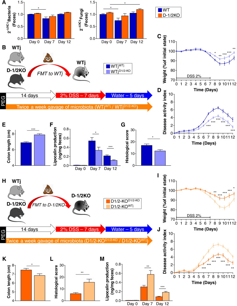
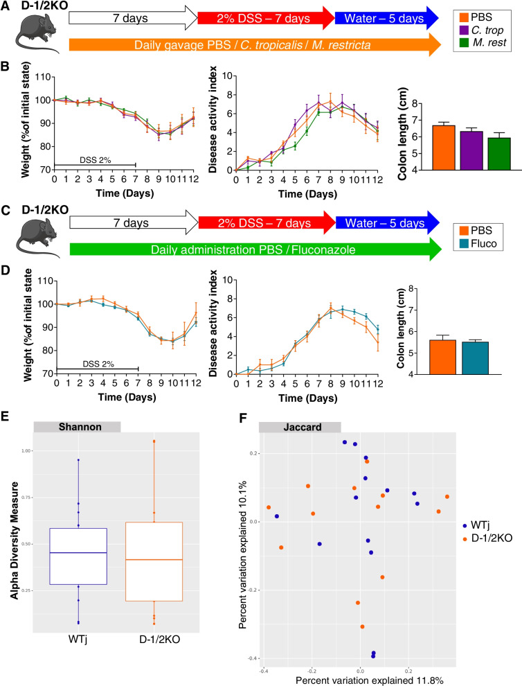
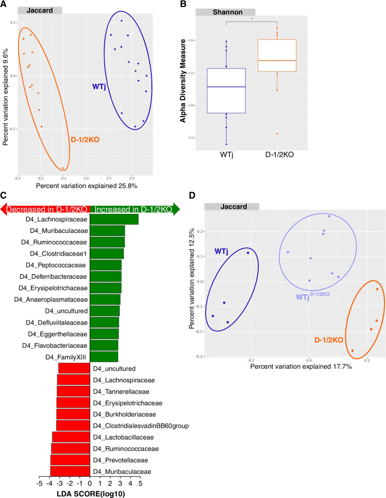
Results: While deletion of Dectin-1 or Dectin-2 did not have a strong effect on DSS-induced colitis, double deletion of Dectin-1 and Dectin-2 significantly protected the mice from colitis. The protection was largely mediated by the gut microbiota, as demonstrated by fecal transfer experiments. Treatment of D-1/2KO mice with opportunistic fungal pathogens or antifungal agents did not affect the protection against gut inflammation, suggesting that the fungal microbiota had no role in the protective phenotype. Amplicon-based microbiota analysis of the fecal bacterial and fungal microbiota of D-1/2KO mice confirmed the absence of changes in the mycobiota but strong modification of the bacterial microbiota. We showed that bacteria from the Lachnospiraceae family were at least partly involved in this protection and that treatment with Blautia hansenii was enough to recapitulate the protection.

Conclusions: Deletion of both the Dectin-1 and Dectin-2 receptors triggered a global shift in the microbial gut environment, affecting, surprisingly, mainly the bacterial population and driving protective effects in colitis. Members of the Lachnospiraceae family seem to play a central role in this protection. These findings provide new insights into the role of the Dectin receptors, which have been described to date as affecting only the fungal population, in intestinal physiopathology and in IBD. Video Abstract.

Keywords: Dectin-1; Dectin-2; Gut inflammation; Immune response; Microbiota.


Conflict of interest statement

The authors declare that they have no competing interests.

Figures

Fig. 1
Fig. 1
Dectin-deficient mice react differently to DSS-induced colitis. A ELISA of the expression of IL-6 (left panel) and TNFα (right panel) in BMDC cultures from wild-type (WT), Dectin-1-deficient (D-1KO), Dectin-2-deficient (D-2KO), and Dectin-1/2-deficient (D-1/2KO) mice stimulated with fungal ligands (zymosan and mannan). BF WT, D-1KO, D-2KO, and D-1/2KO mice received dextran sulfate sodium (DSS) for 7 days. WT n = 13, D-1KO n = 15, D-2KO n = 14, D-1/2KO n = 15. B Experimental design for the administration of DSS. C Weight of DSS-exposed mice. D Disease activity index (DAI) of DSS-exposed mice. E Length of the colons of mice treated with DSS. F Representative H&E-stained images of proximal colon cross sections on day 12 after initial DSS exposure (left panel) and histological scores on day 12 (right panel). G Intestinal inflammation, expressed as the lipocalin levels in feces at day 0, day 7, and day 12. H Intestinal cytokines in the colon (qPCR). I Antimicrobial peptides expressed in the colon (qPCR). For statistical comparisons, (*) indicates D-1/2KO versus WT, and ($) indicates D-2KO versus WT. *p < 0.05, **,$$p < 0.01, ***,$$$p < 0.001
Fig. 2
Fig. 2
The fecal microbiota from double Dectin-deficient mice ameliorates DSS-induced colitis. A Wild-type (WT) and Dectin-1/2-deficient (D-1/2KO) mice received dextran sulfate sodium (DSS) for 7 days. WT n = 13, D-1/2KO n = 15. A Bacterial (left panel) and fungal (right panel) quantities in feces at day 0, day 7, and day 12, determined by qPCR. BG Wild-type mice from Janvier Laboratory transplanted with their own intestinal microbiota (WTjWTj) or transplanted with the intestinal microbiota of Dectin-1/2-deficient mice (WTjD-1/2KO) received DSS for 7 days. WTjWTj n = 18, WTjD-1/2KO n = 20. HM Dectin-1/2-deficient mice transplanted with their own intestinal microbiota (D-1/2KOD-1/2KO) or transplanted with the intestinal microbiota of wild-type mice from Janvier Laboratory (D-1/2KOWTj) received DSS for 7 days. D-1/2KOD-1/2KO n = 17, D-1/2KOWTj n = 18. B/H Experimental design for fecal microbiota transplantation and administration of DSS. C/I Weight of DSS-exposed mice. D/J Disease activity index (DAI) of DSS-exposed mice. E/K Length of the colons of mice treated with DSS. F/L Histological scores on day 12. G/M Intestinal inflammation, expressed as the lipocalin levels in feces at day 0, day 7, and day 12. *p < 0.05, **p < 0.01, ***p < 0.001
Fig. 3
Fig. 3
Fungi do not seem to have an effect on double Dectin-deficient mice with DSS-induced colitis. A, B Dectin-1/2-deficient (D-1/2KO) mice received C. tropicalis (C. trop) or M. restricta (M. rest) and then dextran sulfate sodium (DSS) for 7 days. PBS n = 7, C. trop n = 6, M. rest n = 7. A Experimental design for the administration of fungi and DSS. B Weight of DSS-exposed mice (left panel), disease activity index (DAI) of DSS-exposed mice (middle panel) and length of the colons of mice treated with DSS (right panel). C, D D-1/2KO mice received fluconazole (Fluco) and then DSS for 7 days. PBS n = 3, Fluco n = 8. C Experimental design for the administration of an antifungal agent and DSS. D Weight of DSS-exposed mice (left panel), disease activity index (DAI) of DSS-exposed mice (middle panel) and length of the colons of mice treated with DSS (right panel). E, F Wild-type mice from Janvier Laboratory (WTj) and Dectin-1/2 deficient (D-1/2KO) mice without challenge. WTj n = 14, D-1/2KO n = 13. E Shannon index describing the alpha diversity of the fungal microbiota (ITS) in the fecal microbiota. F Beta diversity. Principal coordinate analysis of Jaccard distance, with each sample colored according to the genotype
Fig. 4
Fig. 4
The bacterial microbiota plays an important role in double Dectin-deficient mouse protection. AC Wild-type mice from Janvier Laboratory (WTj) and Dectin-1/2 deficient (D-1/2KO) mice without challenge. WTj n = 14, D-1/2KO n = 12. A Beta diversity. Principal coordinate analysis of Jaccard distance, with each sample colored according to the genotype. B Shannon index describing the alpha diversity of the bacterial microbiota (16S) in the fecal microbiota. C Taxa with the largest differences (LDA > 2) in abundance by linear discriminant analysis (LEfSe) (LDA > 2). D WTj, D-1/2KO and WTj transplanted with the intestinal microbiota of Dectin-1/2-deficient mice (WTjD-1/2KO) mice without challenge. WTj n = 4, D-1/2KO n = 4, WTjD-1/2KO n = 8. D Beta diversity. Principal coordinate analysis of Jaccard distance, with each sample colored according to the genotype
Fig. 5
Fig. 5
Lachnospiraceae strains can protect mice from DSS-induced colitis. A ELISA of the expression of IL-6 in BMDC cultures from wild-type (WT) and Dectin-1/2-deficient (D-1/2KO) mice stimulated with dead bacteria. BE Mice received several Lachnospiraceae (BM38, BM62, or Blautia) or medium (vehicle) and dextran sulfate sodium (DSS) for 7 days. Vehicle n = 17, BM38 n = 18, BM62 n = 10, Blautia n = 10. B Experimental design for the administration of Lachnospiraceae and DSS. C Weight of DSS-exposed mice. D Disease activity index (DAI) of DSS-exposed mice. E Length of the colons of mice treated with DSS. For statistical comparisons, (*) indicates Blautia versus vehicle, and ($) indicates BM38 versus vehicle. *,$p < 0.05, **p < 0.01, ***p < 0.001

Similar articles

Cited by

References

    1. Liguori G, et al. Fungal dysbiosis in mucosa-associated microbiota of Crohn’s disease patients. J Crohns Colitis. 2016;10(3):296–305. doi: 10.1093/ecco-jcc/jjv209. - DOI - PMC - PubMed
    1. Sokol H, et al. Fungal microbiota dysbiosis in IBD. Gut. 2017;66(6):1039–1048. doi: 10.1136/gutjnl-2015-310746. - DOI - PMC - PubMed
    1. Iliev ID, et al. Interactions between commensal fungi and the C-type lectin receptor Dectin-1 influence colitis. Science. 2012;336(6086):1314–1317. doi: 10.1126/science.1221789. - DOI - PMC - PubMed
    1. Sokol H, et al. Card9 mediates intestinal epithelial cell restitution, T-helper 17 responses, and control of bacterial infection in mice. Gastroenterology. 2013;145(3):591–601 e3. doi: 10.1053/j.gastro.2013.05.047. - DOI - PMC - PubMed
    1. Sovran B, et al. Enterobacteriaceae are essential for the modulation of colitis severity by fungi. Microbiome. 2018;6(1):152. doi: 10.1186/s40168-018-0538-9. - DOI - PMC - PubMed

Publication types

LinkOut - more resources