

Detecting bone lesions in the emergency room with medical infrared thermography
- PMID: 35698224
- PMCID: PMC9190459
- DOI: 10.1186/s12938-022-01005-7
Detecting bone lesions in the emergency room with medical infrared thermography
Abstract
Introduction: Low- to high-energy impact trauma may cause from small fissures up to extended bone losses, which can be classified as closed or opened injuries (when they are visible at a naked eye).
Objective: The aim of this study was to investigate the feasibility of clinical diagnosis of bone trauma through medical infrared thermography, in a hospital emergency room.
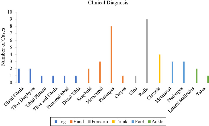
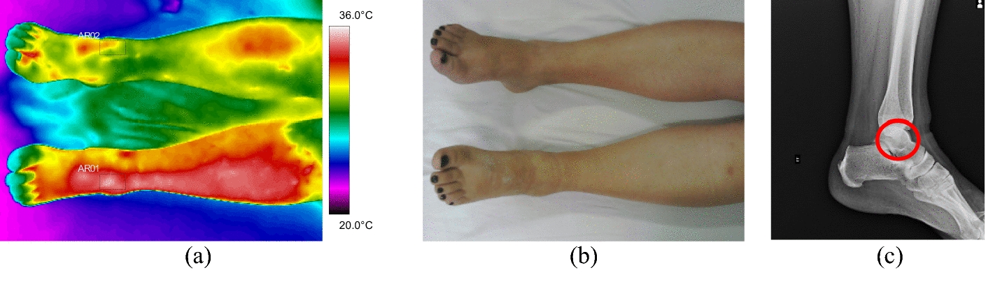
Methods: Forty-five patients with suspected diagnosis of bone fracture were evaluated by means of medical infrared images, and the data correlated with the gold standard radiographic images, in the anteroposterior, lateral, and oblique views, at the orthopedic emergency department. The control group consisted of thermal images of the contralateral reference limb of the volunteers themselves. Data were acquired with a medical grade infrared camera in the regions of interest (ROIs) of leg, hand, forearm, clavicle, foot, and ankle.
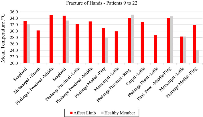
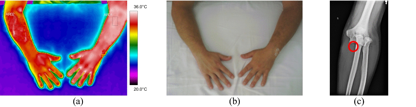
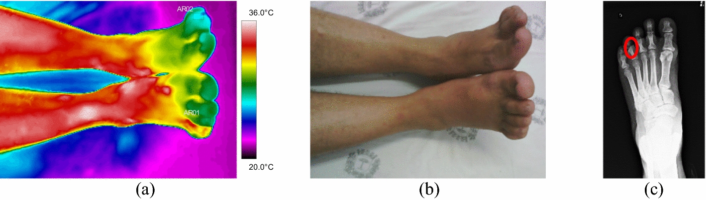
Results: In all patients evaluated with a diagnosis of bone fracture, the mean temperature of the affected limb showed a positive difference greater than 0.9 °C (towards the contralateral), indicating the exact location of the bone trauma according, while the areas diagnosed with reduced blood supply, showed a mean temperature with a negative variation.
Conclusion: Clinical evaluation using infrared imaging indicates a high applicability potential as a tool to support quick diagnosis of bone fractures in patients with acute orthopedic trauma in an emergency medical setting. The thermal results showed important physiological data related to vascularization of the bone fracture and areas adjacent to the trauma well correlated to radiographic examinations.
Keywords: Bone fracture; Bone lesion identification; Diagnostic tool; Emergence room; Infrared thermal imaging; Screening temperature.
© 2022. The Author(s).
Conflict of interest statement
The authors declare no conflict of interest.
Figures


References
-
- Saraiva JA, de Sousa Cabral TK, Mendes JLCF, dos Santos ST, Coelho AG, de Oliveira AM, Torres SG, Cunha FVM. Prevalência de fraturas por acidentes automobilisticos em um hospital público do Piauí. Braz J Health Rev. 2021;4(2):9430–9444. doi: 10.34119/bjhrv4n2-438. - DOI
-
- Schirmer MP. Fraturas por estresse em militares: revisão da literatura fraturas por estresse em militares. 2020.
-
- Corte ACR, Hernandez AJ. Application of medical infrared thermography to sports medicine. Rev Bras Med Esporte. 2016;22:315–319. doi: 10.1590/1517-869220162204160783. - DOI
MeSH terms
Grants and funding
LinkOut - more resources
Full Text Sources
Medical
