

Clinicopathologic profile of gastroenteropancreatic neuroendocrine neoplasms in a referral center of South India
- PMID: 35698638
- PMCID: PMC9187919
Clinicopathologic profile of gastroenteropancreatic neuroendocrine neoplasms in a referral center of South India
Abstract
Background: The neuroendocrine system of the gastroenteropancreatic (GEP) region gives rise to unique, heterogeneous malignancies that need a high index of suspicion to make a diagnosis owing to their indolent course.
Aims: The present study aimed to find the incidence and the differences in the morphologic and immunohistochemical profile of gastroenteropancreatic neuroendocrine tumors (GEPNET) in a referral center of South India, JIPMER, Puducherry, India.
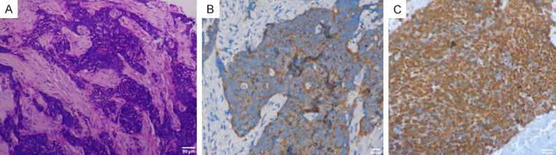
Methods: There were 55 gastroenteropancreatic region neuroendocrine neoplasms (NEN) assessed for demographic, clinical and radiological features. Gross morphological features, histopathological features, mitotic index, Ki67 proliferation index, and immunohistochemical positivity for synaptophysin, chromogranin-A, CD-56, NSE (Neuron Specific Enolase) and pan-cytokeratin (Pan-CK) were also assessed.
Results: The majority were nonfunctional tumors presenting with abdominal pain, gastrointestinal bleed, vomiting, jaundice, and loss of weight and appetite. The sites of involvement according to the order of frequency were duodenum, stomach, rectum, pancreas, ileum, appendix and jejunum. The endoscopic appearance of duodenal and jejunal tumors showed polypoidal, nodular and ulceroproliferative growth. These tumors were diagnosed by preoperative biopsy; 54% of them were grade-1 neuroendocrine tumors exhibiting nesting, trabecular, cord, and solid sheet patterns. All 55 cases were synaptophysin-positive with variable positivity for chromogranin, neuron-specific enolase, CD56, and Pan-CK. Mixed adenoneuroendocrine carcinomas (MANECs) involving the duodenum and stomach comprised 7.3% of all GEPNETs. Pancreatic neuroendocrine tumors constituted 9% of all tumors; one was multifocal. Lymph node metastasis was seen in 12/55 tumors; 6/12 showed liver metastasis also. All metastasizing tumors measured less than 4 cm in size. Statistical correlation of the tumor grade, mitotic count and Ki67 index as analysed by Spearman's correlation between the paired data denoted by rs in 55 tumors showed a strong correlation between mitotic count and Ki67 index; a moderate correlation was noted between the tumor grade and Ki67 index.
Conclusion: The clinicopathologic profile of 55 GEPNET revealed a majority to be sporadic Grade 1 tumor. Tumors that showed lymph node and liver metastasis were less than 4 cm in size. MANECs were found in the duodenum and stomach.
Keywords: Gastroenteropancreatic neuroendocrine tumors; immunohistochemistry; mixed neuroendocrine-non-neuroendocrine neoplasms.
IJCEP Copyright © 2022.
Conflict of interest statement
None.
Figures


References
-
- Møller JE, Pellikka PA, Bernheim AM, Schaff HV, Rubin J, Connolly HM. Prognosis of carcinoid heart disease: analysis of 200 cases over two decades. Circulation. 2005;112:3320–327. - PubMed
-
- Norlén O, Stålberg P, Öberg K, Eriksson J, Hedberg J, Hessman O, Janson ET, Hellman P, Åkerström G. Long-term results of surgery for small intestinal neuroendocrine tumors at a tertiary referral center. World J Surg. 2012;36:1419–1431. - PubMed
-
- Strosberg JR, Weber JM, Feldman M, Coppola D, Meredith K, Kvols LK. Prognostic validity of the American joint committee on cancer staging classification for midgut neuroendocrine tumors. J. Clin. Oncol. 2013;31:420–425. - PubMed
-
- Modlin IM, Oberg K, Chung DC, Jensen RT, de Herder WW, Thakker RV, Caplin M, Delle Fave G, Kaltsas GA, Krenning EP, Moss SF, Nilsson O, Rindi G, Salazar R, Ruszniewski P, Sundin A. Gastroenteropancreatic neuroendocrine tumors. Lancet Oncol. 2008;9:61–72. - PubMed
LinkOut - more resources
Full Text Sources
Research Materials