

Covid-MANet: Multi-task attention network for explainable diagnosis and severity assessment of COVID-19 from CXR images
- PMID: 35698723
- PMCID: PMC9170279
- DOI: 10.1016/j.patcog.2022.108826
Covid-MANet: Multi-task attention network for explainable diagnosis and severity assessment of COVID-19 from CXR images
Abstract
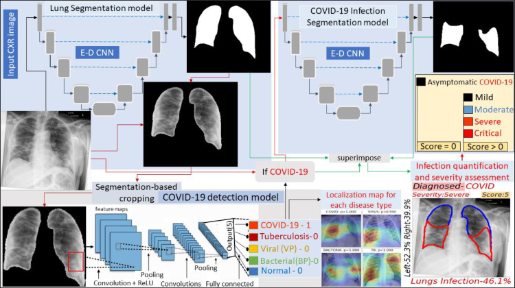
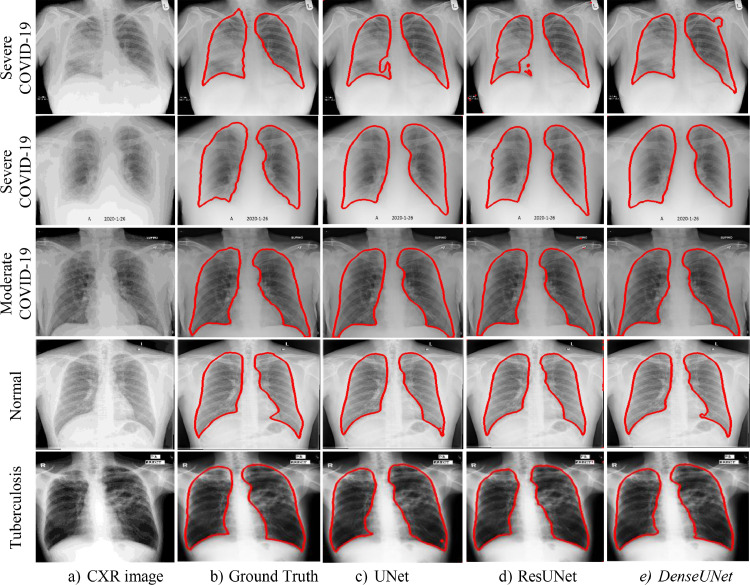
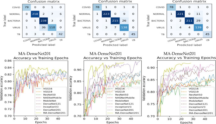
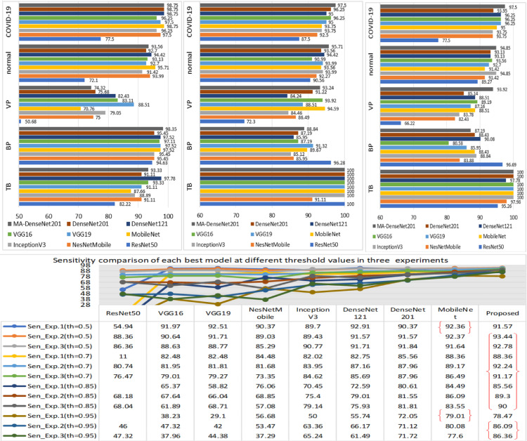
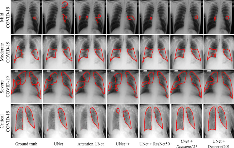
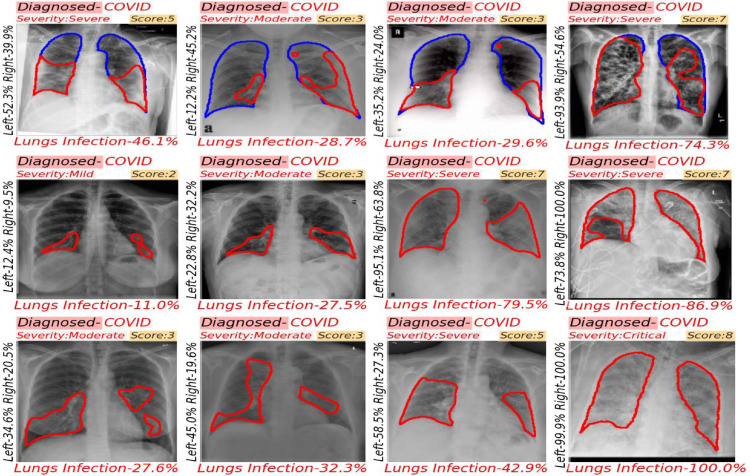
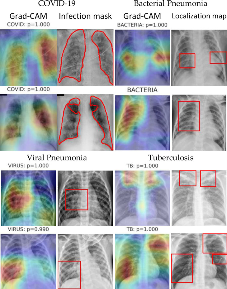
The devastating outbreak of Coronavirus Disease (COVID-19) cases in early 2020 led the world to face health crises. Subsequently, the exponential reproduction rate of COVID-19 disease can only be reduced by early diagnosis of COVID-19 infection cases correctly. The initial research findings reported that radiological examinations using CT and CXR modality have successfully reduced false negatives by RT-PCR test. This research study aims to develop an explainable diagnosis system for the detection and infection region quantification of COVID-19 disease. The existing research studies successfully explored deep learning approaches with higher performance measures but lacked generalization and interpretability for COVID-19 diagnosis. In this study, we address these issues by the Covid-MANet network, an automated end-to-end multi-task attention network that works for 5 classes in three stages for COVID-19 infection screening. The first stage of the Covid-MANet network localizes attention of the model to the relevant lungs region for disease recognition. The second stage of the Covid-MANet network differentiates COVID-19 cases from bacterial pneumonia, viral pneumonia, normal and tuberculosis cases, respectively. To improve the interpretation and explainability, three experiments have been conducted in exploration of the most coherent and appropriate classification approach. Moreover, the multi-scale attention model MA-DenseNet201 proposed for the classification of COVID-19 cases. The final stage of the Covid-MANet network quantifies the proportion of infection and severity of COVID-19 in the lungs. The COVID-19 cases are graded into more specific severity levels such as mild, moderate, severe, and critical as per the score assigned by the RALE scoring system. The MA-DenseNet201 classification model outperforms eight state-of-the-art CNN models, in terms of sensitivity and interpretation with lung localization network. The COVID-19 infection segmentation by UNet with DenseNet121 encoder achieves dice score of 86.15% outperforming UNet, UNet++, AttentionUNet, R2UNet, with VGG16, ResNet50 and DenseNet201 encoder. The proposed network not only classifies images based on the predicted label but also highlights the infection by segmentation/localization of model-focused regions to support explainable decisions. MA-DenseNet201 model with a segmentation-based cropping approach achieves maximum interpretation of 96% with COVID-19 sensitivity of 97.75%. Finally, based on class-varied sensitivity analysis Covid-MANet ensemble network of MA-DenseNet201, ResNet50 and MobileNet achieve 95.05% accuracy and 98.75% COVID-19 sensitivity. The proposed model is externally validated on an unseen dataset, yields 98.17% COVID-19 sensitivity.
Keywords: Chest X-ray; Covid-19; Deep learning; Explainable AI; Infection segmentation; Lung segmentation; Transfer learning.
© 2022 Elsevier Ltd. All rights reserved.
Conflict of interest statement
The authors declare that they have no conflict of interest.
Figures


References
-
- WHO. WHO director-general's opening remarks at the media briefing on COVID-19 - 11 march 2020. https://www.who.int/dg/speeches/detail/who-director-general-s-opening-re... march-2020. 2020
-
- Krizhevsky A., Sutskever I., Hinton G.E. Imagenet classification with deep convolutional neural networks. Adv. Neural Inform. Process. Syst. 2012;25:1097–1105.
LinkOut - more resources
Full Text Sources