Review
. 2022 Jun 24;119(25):429-435.
doi: 10.3238/arztebl.m2022.0231.

Pulmonary and Systemic Pathology in COVID-19—Holistic Pathological Analyses

Affiliations
Review

Pulmonary and Systemic Pathology in COVID-19—Holistic Pathological Analyses

Danny Jonigk et al. Dtsch Arztebl Int. .

Abstract

Background: The COVID-19 pandemic is the third worldwide coronavirus-associated disease outbreak in the past 20 years. Lung involvement, with acute respiratory distress syndrome (ARDS) in severe cases, is the main clinical feature of this disease; the cardiovascular system, the central nervous system, and the gastrointestinal tract can also be affected. The pathophysiology of both pulmonary and extrapulmonary organ damage was almost completely unknown when the pandemic began.

Methods: This review is based on pertinent publications retrieved by a selective search concerning the structural changes and pathophysiology of COVID-19, with a focus on imaging techniques.

Results: Immunohistochemical, electron-microscopic and molecular pathological analyses of tissues obtained by autopsy have improved our understanding of COVID-19 pathophysiology, including molecular regulatory mechanisms. Intussusceptive angiogenesis (IA) has been found to be a prominent pattern of damage in the affected organs of COVID-19 patients. In IA, an existing vessel changes by invagination of the endothelium and formation of an intraluminal septum, ultimately giving rise to two new lumina. This alters hemodynamics within the vessel, leading to a loss of laminar flow and its replacement by turbulent, inhomogeneous flow. IA, which arises because of ischemia due to thrombosis, is itself a risk factor for the generation of further microthrombi; these have been detected in the lungs, heart, liver, kidneys, brain, and placenta of COVID-19 patients.

Conclusion: Studies of autopsy material from various tissues of COVID-19 patients have revealed ultrastructural evidence of altered microvascularity, IA, and multifocal thrombi. These changes may contribute to the pathophysiology of post-acute interstitial fibrotic organ changes as well as to the clinical picture of long COVID.


Figures

Figure 1
Figure 1
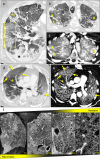
Clinicoradiographic pattern of lung damage in COVID-19 and scaling down to the microscopic level using hierarchical phase-contrast tomography (HIP-CT) a) The COVID-19-associated lung parenchymal changes visualized with high-resolution computed tomography (HRCT) include ground-glass opacities that form, in combination with septal thickening, a so-called crazy paving pattern (square brackets). Consolidations (asterisk) typically only occur with advanced disease. b) HIP-CT allows for stepwise 3D morphological assessment, from the level of clinical CT down to the microscopic scale, and thus enables rescaling of histopathological damage patterns to clinical imaging. See also eFigure 1 for more details. Figure by courtesy of Willi Wagner, Peter Lee, Claire Walsh, and Paul Tafforeau as well as the European Synchrotron Radiation Facility (ESRF) and the University College London (UCL)
Figure 2
Figure 2
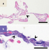
Histological changes in the lungs in COVID-19 over time a) The acute phase is dominated by diffuse alveolar damage (DAD) as a correlate of ARDS in COVID-19. The alveoli are lined by hyaline membranes, which interfere with gas exchange (see arrowhead). b) In addition, minute fibrin thrombi are noted in alveolar capillaries (see arrowheads) in significantly increased numbers. See also eFigure 2 for more details. Magnification: a) 50x, b) 600x Scaling: a) 200 µm, b) 10 µm Figure by courtesy of Jan Christopher Kamp, Mark Kühnel and Lavinia Neubert.
Figure 3
Figure 3
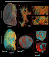
Multi-resolution imaging of a COVID-19 autopsy lung, using hierarchical phase contrast tomography (HiP-CT). Airways are depicted in cyan, the pulmonary vascular structure in red (pulmonary arteries) and yellow (bronchial arteries). Because of the hierarchical nature of HiP-CT, this imaging modality allows identification and segmentation of pulmonary and bronchial circulations from the macroscopic level (a) down to microcirculation (b). In this way, evidence of bronchopulmonary anastomoses and vascular alterations, such as expansion and dilation of the peribronchial plexus and vasa vasorum, can be obtained. Their dysregulation contributes to the typical picture of severe COVID-19 lung involvement. See also eFigure 3 for more details. Figure by courtesy of Claire Walsh, Peter Lee, Claire Walsh, Paul Tafforeau as well as the European Synchrotron Radiation Facility (ESRF) and the University College London (UCL)
Figure 4
Figure 4
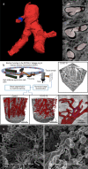
Scanning electron microscope images show changes in alveolar lung architecture in COVID-19 (b) in comparison to the physiological architecture of the lungs (a). Cast preparations of blood vessel, created using the technique of microvascular corrosion casting, show dilated vessels with numerous transluminal tissue columns (arrowheads), the characteristic morphological feature of intussusceptive angiogenesis. See also eFigure 4 and eFigure 5 for more details. Figure by courtesy of Stijn Verleden (Antwerp Surgical Training, Anatomy and Research Centre [ASTARC]), Tim Salditt and Marius Reichhardt (Institute for X-ray Physics, University of Göttingen) a
eFigure 1
eFigure 1
Clinicoradiographic pattern of lung damage in COVID-19 and scaling down to the microscopic level using hierarchical phase-contrast tomography (HIP-CT) a) The COVID-19-associated lung parenchymal changes visualized with high-resolution computed tomography (HRCT) include ground-glass opacities that form, in combination with septal thickening, the so-called crazy paving pattern ([]). Consolidations (marked with asterisk) typically only occur with advanced disease. Perfusion deficits may occur in parts of the lung with or without radiographically identifiable parenchymal changes. b–e) Similarly, relative hyperperfusion may be demonstrable in normally ventilated parts of the lung (marked by yellow crosses in b and c) and also in areas of ground-glass attenuation (arrows in d and e; the plus sign indicates normally ventilated areas in d and e);these radiologic changes indicate microvascular dysfunction. f) HIP-CT allows for stepwise 3D morphological assessment, from the level of clinical CT (Fi) down to the microscopic scale (Fiiii), and thus enables rescaling of histopathological damage patterns to clinical imaging. Figure by courtesy of Willi Wagner, Peter Lee, Claire Walsh, and Paul Tafforeau as well as the European Synchrotron Radiation Facility (ESRF) and the University College London (UCL)
eFigure 2
eFigure 2
Histological and molecular pathology changes in the lungs in COVID-19 over time Magnification: a) 50x , b) 600x c) 100x, d) 50x. Scaling: a) 200 µm, b) 10 µm, c) 20 µm, d) 200 µm a) The acute phase is dominated by diffuse alveolar damage (DAD) as a correlate of ARDS in COVID-19. The alveoli are lined by hyaline membranes, which interfere with gas exchange (see arrowhead). b) In addition, minute fibrin thrombi are noted in alveolar capillaries (see arrowheads) in significantly increased numbers. c) Fibrin thrombi are found approx. 10 times more frequently compared to viral pneumonias, such as severe influenza. Immunohistochemistry reveals viral proteins (see here spike protein in brown) of SARS-CoV-2, especially in macrophages, alveolar epithelial cells (type I and type II), and in endothelial cells (see arrowhead). Direct infection of endothelial cells by SARS-CoV-2 triggers angiocentric inflammation. d) In the further course of the disease, the histological picture is dominated by fibrotic remodeling. Extensive widening of alveolar septa, combined with the pattern of organizing pneumonia with intra-alveolar mesenchymal sprouting of the parenchyma (see arrow) as well as residues of diffuse alveolar damage with hyaline membranes even long after the end of the acute phase (see arrowhead) are frequently seen. e) Schematic representation of the results of a gene expression analysis of over 700 genes, using nanostring encounter systems on lung parenchyma of patients who died of COVID-19. A comparison of patients who died within the first week of hospitalization with patients who died later than 10 days of hospitalization (median: 14 days) found marked inflammation along with initial fibrotic activity in the group of patients who died earlier, whereas this ratio was reversed in the late group. Molecular signaling pathways associated with fibrosis and inflammation are shown in blue and red, respectively. Other significantly regulated signaling pathways were summarized under “other” (gray). Figure adapted from (e20) Figure by courtesy of Jan Christopher Kamp, Mark Kühnel and Lavinia Neubert
eFigure 3
eFigure 3
Multi-resolution imaging of a COVID-19 autopsy lung, using hierarchical phase contrast tomography (HiP-CT). Airways are depicted in cyan, the pulmonary vascular structure in red (pulmonary arteries) and yellow (bronchial arteries). In a) to d), both the airways and the vascular architecture of the lung are depicted. Thanks to the hierarchical nature of HiP-CT, this imaging modality allows identification and segmentation of pulmonary and bronchial circulations from the macroscopic level (a) down to microcirculation (b). In this way, evidence of bronchopulmonary anastomoses and vascular alterations, such as expansion and dilation of the peribronchial plexus and vasa vasorum, can be obtained. Their dysregulation contributes to the typical picture of severe COVID-19 lung involvement. Airway segmentation (from the bronchi to the alveoli proper) across the entire lobe of the lung in e) to h) highlights the segmental distribution between severely affected and largely spared areas of the COVID-19-related damage. The color scale shows the proportion of open air space. High-resolution visualization of an interlobular septum in f) reveals areas with more pronounced fibrotic remodeling immediately adjacent to less damaged areas. Thanks to HiP-CT, these distinct areas could be related to the anatomical structure of the so-called secondary pulmonary lobule. Visualizations in even higher resolutions in g) and h) show differences in airway morphology from the large bronchi down to the individual alveoli. Figure by courtesy of Claire Walsh, Peter Lee, Paul Tafforeau as well as the European Synchrotron Radiation Facility (ESRF) and the University College London (UCL)
eFigure 4
eFigure 4
a) 3D-micro-CT reconstruction to show an obliterated blood vessel. A reconstruction of the vascular tree, based on serial micro-CT images, revealed a ramified vessel (*) with complete obliteration (blue) and opening further distally (red). In the remainder of the vascular tree (below the asterisk in light pink), no signs of vascular obliteration are found. b) Three-dimensional visualization of small tissue samples, using synchrotron radiation. In the upper right corner of the image, a reconstruction of heart tissue (volume approximately 0.3 mm edge length) is shown as an example. In the lower series, large volumes (diameter of 1 mm) are shown: for COVID-19 and for a control. In comparison to the normal heart, the heart of the COVID-19 patient shows a chaotically remodeled network with an abundance of splits, branches and loops. These changes represent the first direct visual evidence of intussusceptive angiogenesis, a key driver of tissue damage in COVID-19. In order to visualize the capillary network, the vessels were “identified” in the three-dimensionally reconstructed volume, initially with the help of machine learning, and finally mathematically analyzed and quantified using graphs. c) Scanning electron microscope images reveal changes in alveolar lung architecture in COVID-19 (image on the right) in comparison to the normal architecture of the lungs (image on the left). Cast preparations of blood vessel, created using the technique of microvascular corrosion casting, show dilated vessels with numerous transluminal tissue columns (see arrowheads), the characteristic morphological feature of intussusceptive angiogenesis. Figure by courtesy of Stijn Verleden (Antwerp Surgical Training, Anatomy and Research Centre [ASTARC]), Tim Salditt and Marius Reichhardt (Institute for X-ray Physics, University of Göttingen)
eFigure 5
eFigure 5
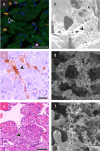
COVID-19 in other organs a, b) COVID-19 manifestation in the heart: Multiplex imaging of heart muscle tissue (a) of a patient who died of COVID-19, with prominent macrophage infiltration (CD68, yellow, see arrowhead) as well as sporadic lymphocytes (B cell, CD20, in purple). Tie-2-positive macrophages are found significantly increased in heart tissue of COVID-19 patients, while classic signs of myocarditis, such as prominent lymphocytic infiltrates and muscle necrosis, are missing. Corrosion casts of the capillary bed (b) reveal an increased number of intussusceptive pillars/tissue columns (see arrowhead) as an expression of increased intussusceptive neoangiogenesis as well as an increased number of thrombi in the smallest capillaries. c, d) COVID-19 manifestation in the liver: Immunohistochemical staining for activated fibrin (c) reveals multiple thrombi within the hepatic sinus (see arrowhead). The scanning electron microscope image in d) shows systemic vascular changes of the capillary bed in COVID-19. e, f) COVID-19 manifestation in the placenta. Conventional morphology reveals lymphocytic placentitis with placental infarction (see arrowhead). In corrosion casts of the capillary bed (f), an increased number of intussusceptive pillars as an expression of increased intussusceptive neoangiogenesis is revealed, which is in line with the lung and heart findings. Scaling: a) 100 µm, c) 20 µm, e): 100 µm. Figure by courtesy of Kerstin Bahr (University of Mainz) und Detlef Schuppan (University of Mainz), Lavinia Neubert, Mark Kühnel, and Peter Braubach (Hannover Medical School)

References

    1. Zhu N, Zhang D, Wang W, et al. A novel coronavirus from patients with pneumonia in China, 2019. N Engl J Med. 2020;382:727–733. - PMC - PubMed
    1. Kommoss FKF, Schwab C, Tavernar L, et al. The pathology of severe COVID-19 related lung damage— mechanistic and therapeutic implications. Dtsch Arztebl Int. 2020;117:500–506. - PMC - PubMed
    1. Ackermann M, Werlein C, Länger F, Kühnel MP, Jonigk DD. COVID-19: effects on the lungs and heart. Pathologe. 2021;42:164–171. - PMC - PubMed
    1. Ritschel N, Radbruch H, Herden C, et al. COVID-19 and the central and peripheral nervous system. Pathologe. 2021;42:172–182. - PMC - PubMed
    1. Guo M, Tao W, Flavell RA, Zhu S. Potential intestinal infection and faecal-oral transmission of SARS-CoV-2. Nat Rev Gastroenterol Hepatol. 2021;18:269–283. - PMC - PubMed