. 2022 Jul;36(4):1491-1501.
doi: 10.1111/jvim.16447. Epub 2022 Jun 14.

Effects of intravenous administration of peripheral blood-derived mesenchymal stromal cells after infusion of lipopolysaccharide in horses

Affiliations

Effects of intravenous administration of peripheral blood-derived mesenchymal stromal cells after infusion of lipopolysaccharide in horses

Sandra D Taylor et al. J Vet Intern Med. 2022 Jul.

Abstract

Background: A systemic and dysregulated immune response to infection contributes to morbidity and mortality associated with sepsis. Peripheral blood-derived mesenchymal stromal cells (PB-MSC) mitigate inflammation in animal models of sepsis. Allogeneic PB-MSC administered IV to horses is well-tolerated but therapeutic benefits are unknown.

Hypothesis: After IV lipopolysaccharide (LPS) infusion, horses treated with PB-MSC would have less severe clinical signs, clinicopathological abnormalities, inflammatory cytokine gene expression, and oxidative stress compared to controls administered a placebo.

Animals: Sixteen horses were included in this study.

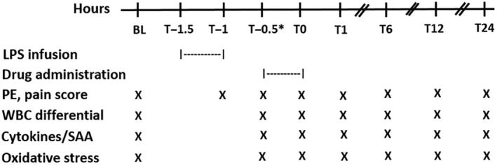
Methods: A randomized placebo-controlled experimental trial was performed. Sixteen healthy horses were assigned to 1 of 2 treatment groups (1 × 109 PB-MSC or saline placebo). Treatments were administered 30 minutes after completion of LPS infusion of approximately 30 ng/kg. Clinical signs, clinicopathological variables, inflammatory cytokine gene expression, and oxidative stress markers were assessed at various time points over a 24-hour period.

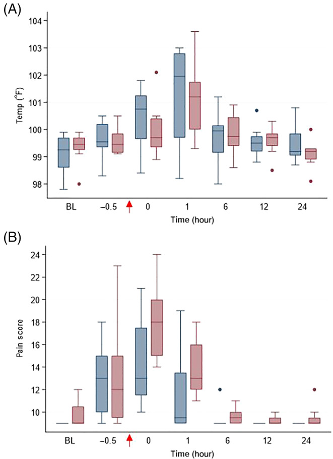
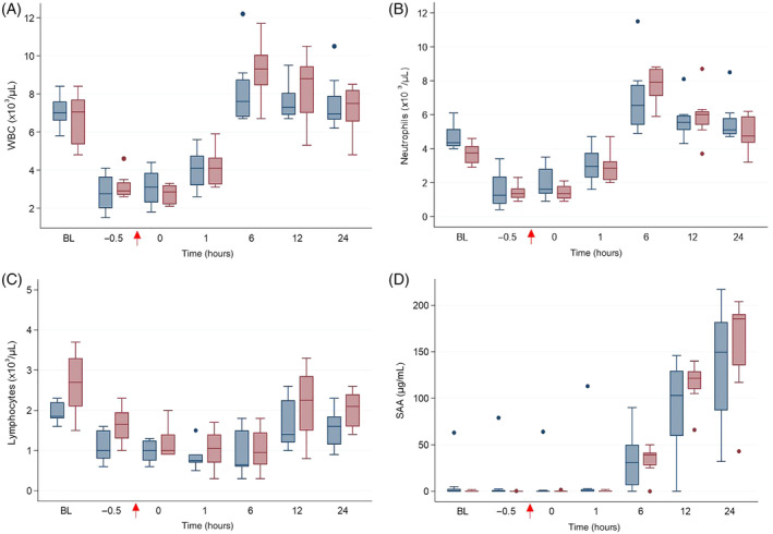
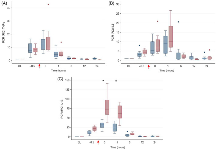
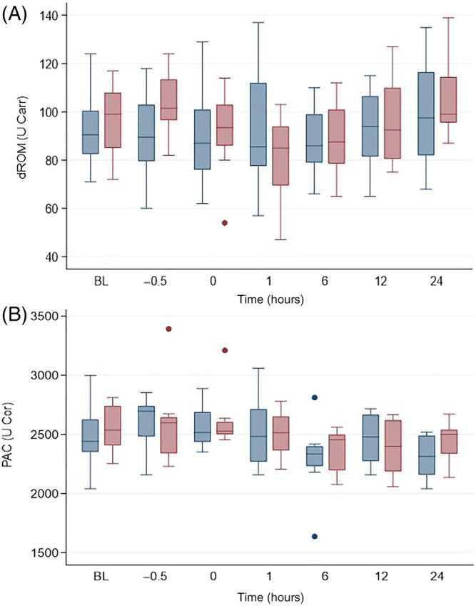
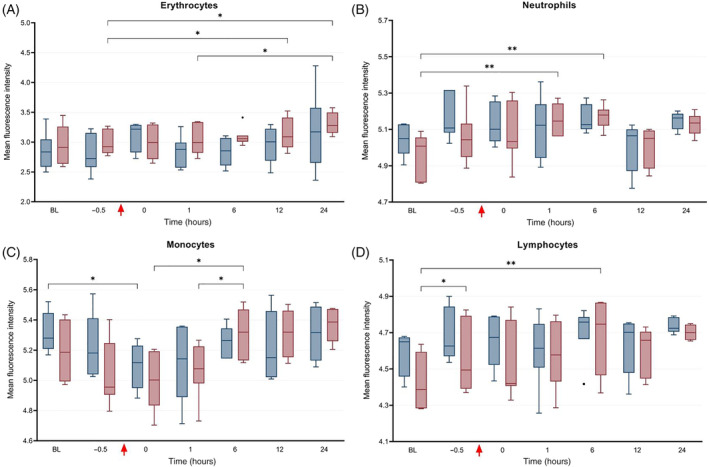
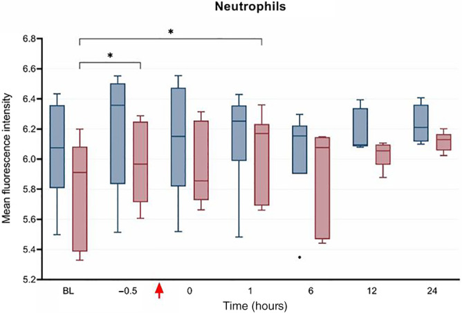
Results: A predictable response to IV LPS infusion was observed in all horses. At the dose administered, there was no significant effect of PB-MSC on clinical signs, clinicopathological variables, or inflammatory cytokine gene expression at any time point. Antioxidant potential was not different between treatment groups, but intracellular ROS increased over time in the placebo group. Other variables that changed over time were likely due to effects of IV LPS infusion.

Conclusions and clinical importance: Administration of allogeneic PB-MSC did not cause clinically detectable adverse effects in healthy horses. The dose of PB-MSC used here is unlikely to exert a beneficial effect in endotoxemic horses.

Keywords: allogeneic; endotoxemia; horses; oxidative stress.


Conflict of interest statement

George E. Moore serves as Consulting Editor for Experimental Design and Statistics for the Journal of Veterinary Internal Medicine. He was not involved in review of this manuscript. No other authors have a conflict of interest.

Figures

FIGURE 1
FIGURE 1
Daily schedule from baseline (BL) to 24 hours after drug administration in 16 adult horses. The asterisk indicates that the T−0.5 blood sample was collected immediately before drug administration. LPS, lipopolysaccharide; PE, physical examination; WBC, white blood cell; SAA, serum amyloid A
FIGURE 2
FIGURE 2
(A) Rectal temperature (°F) and (B) pain score in horses administered peripheral blood‐derived mesenchymal stromal cells (PB‐MSC; n = 8, blue bars) or saline (placebo; n = 8; red bars) after LPS infusion. The arrow indicates completion time of drug administration (PB‐MSC or saline). Box and whisker plots are displayed with median (line) within box (IQR). Whiskers represent range except for points, which extend >1.5 times IQR; these points or outliers are displayed as filled circles. BL: baseline
FIGURE 3
FIGURE 3
(A) WBC count, (B) segmented neutrophil count, (C) lymphocyte count and (D) serum amyloid A (SAA) in horses administered peripheral blood‐derived mesenchymal stromal cells (PB‐MSC; n = 8, blue bars) or saline (placebo; n = 8; red bars) after LPS infusion. The arrow indicates completion time of drug administration (PB‐MSC or saline). Box and whisker plots are displayed with median (line) within box (IQR). Whiskers represent range except for points, which extend >1.5 times IQR; these points or outliers are displayed as filled circles. BL: baseline
FIGURE 4
FIGURE 4
Gene expression of (A) TNFα, (B) ILprio6, and (C) IL1β in horses administered peripheral blood‐derived mesenchymal stromal cells (PB‐MSC; n = 8, blue bars) or saline (placebo; n = 8; red bars) after LPS infusion. The arrow indicates completion time of drug administration (PB‐MSC or saline). Box and whisker plots are displayed with median (line) within box (IQR). Whiskers represent range except for points, which extend >1.5 times IQR; these points or outliers are displayed as filled circles. Asterisks denote differences between treatment groups. BL: baseline
FIGURE 5
FIGURE 5
Determinants of (A) reactive oxygen metabolites (dROM) and (B) plasma antioxidant capacity (PAC) in horses administered peripheral blood‐derived mesenchymal stromal cells (PB‐MSC; n = 8, blue bars) or saline (placebo; n = 8; red bars) after LPS infusion. The arrow indicates completion time of drug administration (PB‐MSC or saline). Box and whisker plots are displayed with median (line) within box (IQR). Whiskers represent range except for points, which extend >1.5 times IQR; these points or outliers are displayed as filled circles. BL: baseline
FIGURE 6
FIGURE 6
Median fluorescence intensity (IQR and range) as determined by flow cytometry representing the direct concentration of intracellular reactive oxygen species (ie, current oxidative status) in peripheral blood (A) erythrocytes, (B) neutrophils, (C) monocytes, and (D) lymphocytes in horses administered peripheral blood‐derived mesenchymal stromal cells (PB‐MSC; erythrocytes: n = 8, leukocytes: n = 6; red bars) or saline (placebo; n = 7, blue bars) after LPS infusion. The bars indicate differences between groups. *P < .05; **P < .01
FIGURE 7
FIGURE 7
Median fluorescence intensity (IQR and range) as determined by flow cytometry representing the antioxidant potential (ie, intracellular concentration of reactive oxygen species after stimulation with hydrogen peroxide) in neutrophils from horses administered peripheral blood‐derived mesenchymal stromal cells (PB‐MSC: n = 6, red bars) or saline (placebo; n = 7, blue bars) after LPS infusion. The bars indicate differences between groups. *P < .05

Similar articles

Cited by

References

    1. Cohen ND. Causes of and farm management factors associated with disease and death in foals. J Am Vet Med Assoc. 1994;204:1644‐1651. - PubMed
    1. Peek SF, Semrad S, McGuirk SM, et al. Prognostic value of clinicopathologic variables obtained at admission and effect of antiendotoxin plasma on survival in septic and critically ill foals. J Vet Intern Med. 2006;20:569‐574. - PubMed
    1. Arroyo MG, Slovis NM, Moore GE, Taylor SD. Factors associated with survival in 97 horses with septic pleuropneumonia. J Vet Intern Med. 2017;31:894‐900. - PMC - PubMed
    1. Bone RC, Balk RA, Cerra FB, et al. Definitions for sepsis and organ failure and guidelines for the use of innovative therapies in sepsis. The ACCP/SCCM consensus conference Committee. American College of Chest Physicians/Society of Critical Care Medicine. Chest. 1992;101:1644‐1655. - PubMed
    1. Werners AH, Bull S, Fink‐Gremmels J. Endotoxaemia: a review with implications for the horse. Equine Vet J. 2005;37:371‐383. - PubMed

Publication types