. 2022 Jul 9;35(2):ivac166.
doi: 10.1093/icvts/ivac166.

Strategy for lung parenchyma-sparing bronchial resection: a case series report

Affiliations

Strategy for lung parenchyma-sparing bronchial resection: a case series report

Akihiro Ohsumi et al. Interact Cardiovasc Thorac Surg. .

Abstract

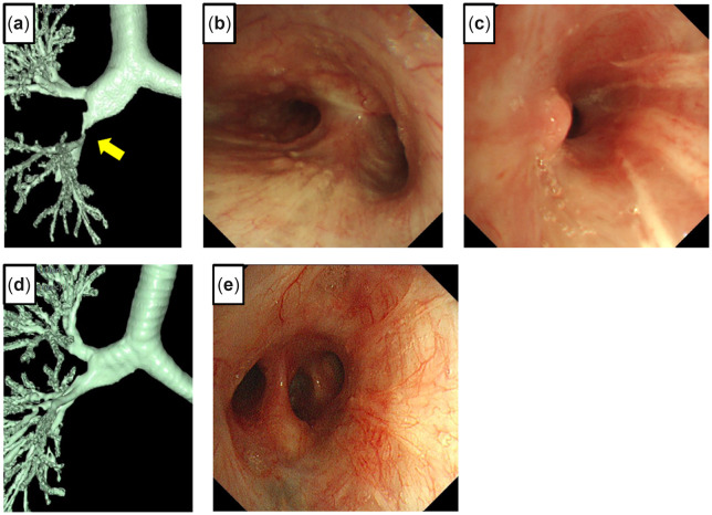
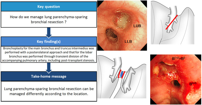
Lung parenchyma-sparing bronchial resection is uncommon, and the operative procedure depends on the cause and location of the stenosis. We present 6 cases and discuss the different surgical strategies for sleeve resection of the central airway without lung resection. Bronchoplasty for the main bronchus and truncus intermedius was performed with a posterolateral approach. We resected the right main bronchus including the right lateral wall of the lower trachea and half of the carina obliquely and performed an anastomosis. The tumour in the left lobar bronchus was exposed and removed by transient division of the accompanying pulmonary artery. Although post-transplant stenosis and malacia can pose a challenge, bronchoplasty can be used as a definitive treatment in experienced centres.

Keywords: Bronchial sleeve resection; Bronchoplasty; Lung cancer; Lung parenchyma-sparing.


Figures

Figure 1:
Figure 1:
Chest computed tomography shows a tumour occupying the lower left main bronchus (arrow points to the tumour) (a). A 3-dimensional computed tomography scan shows almost total obstruction by the tumour (arrow points to the obstruction) (b). Bronchoscopy shows a tumour obstructing the lower left main bronchus (c). Postoperative 3-dimensional computed tomography shows airway lumen patency (d).
Figure 2:
Figure 2:
Chest computed tomography in a coronal view shows a tumour around the proximal right main bronchus (a). Bronchoscopy shows the membranous part of the right lateral wall of the lower trachea pressed by a tumour (b). The bronchus occupied by a tumour was divided obliquely at the proximal and distal sides (c). A 3-dimensional computed tomography scan shows airway lumen patency of the anastomosis (d). Bronchoscopy 2 months after the operation shows healing of the anastomosis (truncus intermedius).
Figure 3:
Figure 3:
Three-dimensional computed tomography shows severe stricture (arrow points to the stricture) (a). Bronchoscopy shows a pinhole with granulated tissue at the orifice of the right main bronchus (b). A 3-dimensional scan shows decent passage of the anastomosis (c). Bronchoscopy 3 months after surgery shows healing and lumen patency of the anastomosis (d).
Figure 4:
Figure 4:
Chest computed tomography shows a tumour occupying the left upper bronchus (LUB); the arrow points to a tumour (a). Bronchoscopy before surgery shows a tumour obstructing the LUB (LLL: left lower bronchus) (b). The interlobar pulmonary artery was divided obliquely to expose the LUB (c). The LUB was exposed and removed with the tumour (d).
Figure 5:
Figure 5:
Three-dimensional computed tomography 6 months after a lung transplant shows severe stricture of the truncus intermedius (TIM) (a). Bronchoscopy 6 months after the lung transplant shows healing with complete lumen patency of the right main bronchial anastomosis (b) and severe stricture at the distal side of the TIM (c). A 3-dimensional computed tomography scan 2 years after TIM reconstruction shows lumen patency (d). Bronchoscopy 2 years after reconstruction shows healing of the anastomosis (e).
Figure 6:
Figure 6:
Chest computed tomography shows kinking of the anastomosed right upper bronchus (RUB) (a). Bronchoscopy before rethoracotomy shows narrowing of the anastomosed RUB (b). A chest computed tomography scan shows passage of the re-anastomosed RUB (c). Bronchoscopy shows healing and passage of the re-anastomosed RUB (d).
None

References

    1. Hamada S, Ohsumi A, Goto T, Hamaji M, Date H, Hirai T.. Delayed right main bronchus stenosis with involvement of the tracheal carina after blunt chest trauma: successful treatment with tracheobronchial anastomosis. Arch Bronconeumol 2020; S0300-2896(20)30341-0.[TQ1] - PubMed
    1. Lucchi M, Melfi F, Ribechini A, Dini P, Duranti L, Fontanini G. et al. Sleeve and wedge parenchyma-sparing bronchial resections in low-grade neoplasms of the bronchial airway. J Thorac Cardiovasc Surg 2007;134:373–7. - PubMed
    1. Stamatis G, Fechner S, Rocha M, Weinreich G.. Resection of the tracheobronchial bifurcation with complete preservation of lung parenchyma. Ann Thorac Surg 2017;104:1741–7. - PubMed
    1. Dell'Amore A, Chen L, Monaci N, Campisi A, Wang Z, Mammana M. et al. Total lung-sparing surgery for tracheobronchial low-grade malignancies. Ann Thorac Surg 2021;112:450–8. - PubMed
    1. Newton JR Jr, Grillo HC, Mathise DJ.. Main bronchial sleeve resection with pulmonary conservation. Ann Thorac Surg 1991;52:1272–80. - PubMed

LinkOut - more resources