Multicenter Study
. 2022 Oct;305(1):68-79.
doi: 10.1148/radiol.212929. Epub 2022 Jun 14.

Validation of Artificial Intelligence Cardiac MRI Measurements: Relationship to Heart Catheterization and Mortality Prediction

Affiliations
Multicenter Study

Validation of Artificial Intelligence Cardiac MRI Measurements: Relationship to Heart Catheterization and Mortality Prediction

Samer Alabed et al. Radiology. 2022 Oct.

Erratum in

Abstract

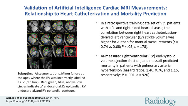
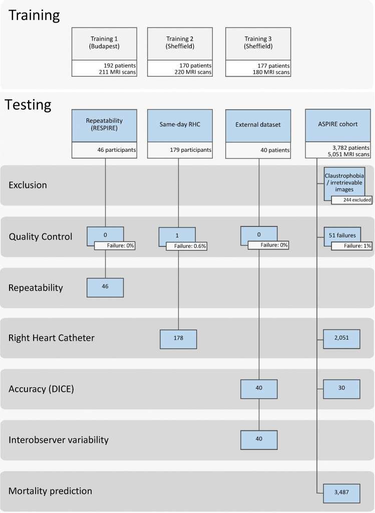
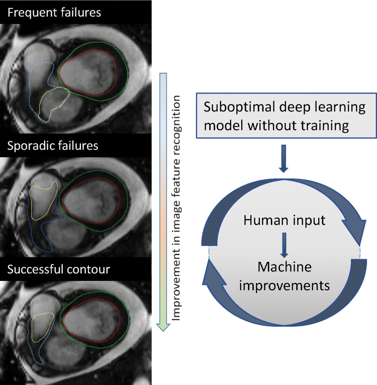
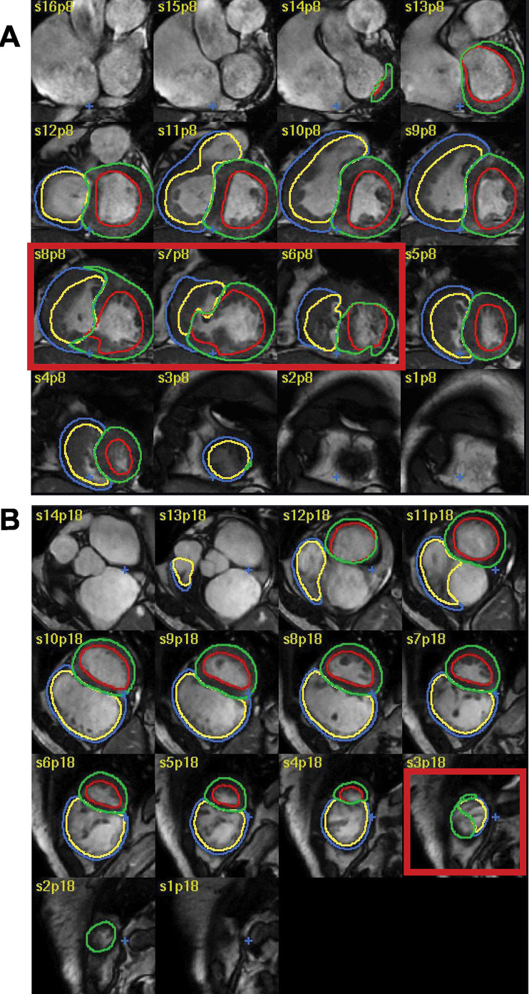
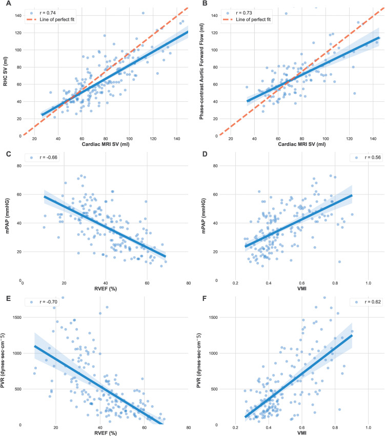
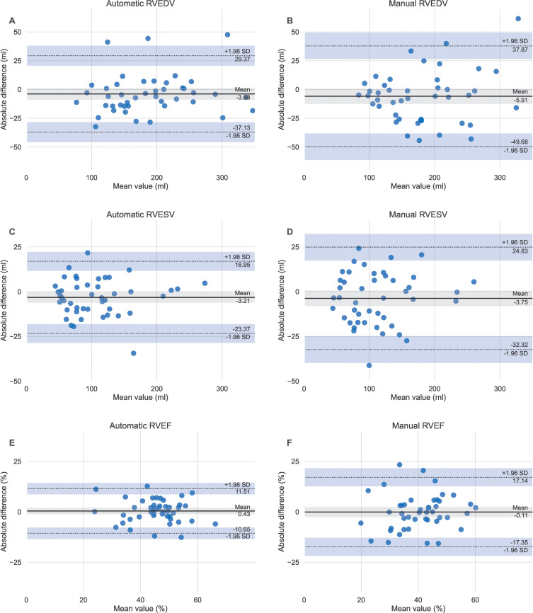
Background Cardiac MRI measurements have diagnostic and prognostic value in the evaluation of cardiopulmonary disease. Artificial intelligence approaches to automate cardiac MRI segmentation are emerging but require clinical testing. Purpose To develop and evaluate a deep learning tool for quantitative evaluation of cardiac MRI functional studies and assess its use for prognosis in patients suspected of having pulmonary hypertension. Materials and Methods A retrospective multicenter and multivendor data set was used to develop a deep learning-based cardiac MRI contouring model using a cohort of patients suspected of having cardiopulmonary disease from multiple pathologic causes. Correlation with same-day right heart catheterization (RHC) and scan-rescan repeatability was assessed in prospectively recruited participants. Prognostic impact was assessed using Cox proportional hazard regression analysis of 3487 patients from the ASPIRE (Assessing the Severity of Pulmonary Hypertension In a Pulmonary Hypertension Referral Centre) registry, including a subset of 920 patients with pulmonary arterial hypertension. The generalizability of the automatic assessment was evaluated in 40 multivendor studies from 32 centers. Results The training data set included 539 patients (mean age, 54 years ± 20 [SD]; 315 women). Automatic cardiac MRI measurements were better correlated with RHC parameters than were manual measurements, including left ventricular stroke volume (r = 0.72 vs 0.68; P = .03). Interstudy repeatability of cardiac MRI measurements was high for all automatic measurements (intraclass correlation coefficient range, 0.79-0.99) and similarly repeatable to manual measurements (all paired t test P > .05). Automated right ventricle and left ventricle cardiac MRI measurements were associated with mortality in patients suspected of having pulmonary hypertension. Conclusion An automatic cardiac MRI measurement approach was developed and tested in a large cohort of patients, including a broad spectrum of right ventricular and left ventricular conditions, with internal and external testing. Fully automatic cardiac MRI assessment correlated strongly with invasive hemodynamics, had prognostic value, were highly repeatable, and showed excellent generalizability. Clinical trial registration no. NCT03841344 Published under a CC BY 4.0 license. Online supplemental material is available for this article. See also the editorial by Ambale-Venkatesh and Lima in this issue. An earlier incorrect version appeared online. This article was corrected on June 27, 2022.


Conflict of interest statement

Disclosures of conflicts of interest: S.A. No relevant relationships. F.A. No relevant relationships. K.D. Research study funding from Janssen Pharmaceuticals. K.K. No relevant relationships. M.S. No relevant relationships. P.G. No relevant relationships. P.J.H.d.K. No relevant relationships. A.T. No relevant relationships. Y.S. No relevant relationships. C.J. No relevant relationships. M.M. No relevant relationships. S.S. No relevant relationships. D.C. No relevant relationships. S.W. Funding from Janssen Pharmaceuticals. P.M. No relevant relationships. A.M.K.R. No relevant relationships. R.C. Honoraria for speaking and advisory boards from Janssen and MSD. N.H. Payment for lectures from MSD, Janssen; participation on a DataSafety Monitoring Board or Advisory Board from MSD, Janssen. J.M.W. No relevant relationships. D.P.O. No relevant relationships. H.L. No relevant relationships. D.G.K. Grant funding from Janssen, GSK; consulting fees from Acceleron, Janssen, GSK, MSD, Ferrer; payments for lectures from Janssen, GSK, MSD, Ferrer; medicolegal fees from Slater and Gordon and Irwin Mitchell; support for meetings/travel from Janssen, MSD; participation on a DataSafety Monitoring Board or Advisory Board from Acceleron, Janssen, GSK; chair of UK National Audit and member of the Clinical Reference Group for Pulmonary Hypertension. R.J.v.d.G. No relevant relationships. A.J.S. Consulting fees from Janssen Pharmaceuticals, GE; payment for lectures from Janssen Pharmaceuticals; support for attending meetings/travel from Janssen Pharmaceuticals; patent for medical image processing.

Figures

None
Graphical abstract
Study participant flow chart for the training and testing cohorts.
ASPIRE = Assessing the Severity of Pulmonary Hypertension In a Pulmonary
Hypertension Referral Centre, RESPIRE = Repeatability and Sensitivity to
Change of Noninvasive End Points in Pulmonary Arterial Hypertension, RHC 5
right heart catheter.
Figure 1:
Study participant flow chart for the training and testing cohorts. ASPIRE = Assessing the Severity of Pulmonary Hypertension In a Pulmonary Hypertension Referral Centre, RESPIRE = Repeatability and Sensitivity to Change of Noninvasive End Points in Pulmonary Arterial Hypertension, RHC 5 right heart catheter.
Example of improvement following additional training. This example
demonstrates improvement of the right ventricular base after additional
training. The first model missed the right ventricular outflow tract and
included the right atrium instead (top image: yellow annotation showing
right ventricular endocardial border), whereas the final model correctly
included the right ventricular outflow tract and excluded the right atrium
(bottom image).
Figure 2:
Example of improvement following additional training. This example demonstrates improvement of the right ventricular base after additional training. The first model missed the right ventricular outflow tract and included the right atrium instead (top image: yellow annotation showing right ventricular endocardial border), whereas the final model correctly included the right ventricular outflow tract and excluded the right atrium (bottom image).
Examples of failed and suboptimal artificial intelligence (AI)
segmentations. (A) Major failure because of congenital heart disease causing
the left ventricular (LV) contours to extend into the right ventricle (RV;
red box). (B) Minor failure at the apex where the RV was incorrectly
labelled as LV (red box). The red, green, blue, and yellow circles indicate
the LV endocardial, LV epicardial, RV endocardial, and RV epicardial
contors, respectively.
Figure 3:
Examples of failed and suboptimal artificial intelligence (AI) segmentations. (A) Major failure because of congenital heart disease causing the left ventricular (LV) contours to extend into the right ventricle (RV; red box). (B) Minor failure at the apex where the RV was incorrectly labelled as LV (red box). The red, green, blue, and yellow circles indicate the LV endocardial, LV epicardial, RV endocardial, and RV epicardial contors, respectively.
Graphs show the relationship between automatic cardiac MRI
measurements, right heart catheterization (RHC) and phase-contrast aortic
flow. Automatic cardiac MRI measurements were compared to (A) RHC stroke
volume (SV) and (B) phase-contrast aortic flow in 178 patients of the
same-day RHC cohort. (C) Mean pulmonary artery pressure (mPAP) was compared
with right ventricle ejection fraction (RVEF) and (D) ventricular mass index
(VMI; RV mass–to–LV mass). (E) Pulmonary vascular resistance
(PVR) was compared to RVEF and (F) VMI.
Figure 4:
Graphs show the relationship between automatic cardiac MRI measurements, right heart catheterization (RHC) and phase-contrast aortic flow. Automatic cardiac MRI measurements were compared to (A) RHC stroke volume (SV) and (B) phase-contrast aortic flow in 178 patients of the same-day RHC cohort. (C) Mean pulmonary artery pressure (mPAP) was compared with right ventricle ejection fraction (RVEF) and (D) ventricular mass index (VMI; RV mass–to–LV mass). (E) Pulmonary vascular resistance (PVR) was compared to RVEF and (F) VMI.
Bland-Altman plots of scan-rescan repeatability for the automatic
compared to the manual right ventricular parameters. Same day scan-rescan
cardiac MRIs were performed in 46 participants to compare the repeatability
of the (A, C, D) automatic and (B, D, F) manual measurements. RVEDV = right
ventricular end-diastolic volume, RVEF = right ventricular ejection
fraction, RVESV = right ventricular end-systolic volume.
Figure 5:
Bland-Altman plots of scan-rescan repeatability for the automatic compared to the manual right ventricular parameters. Same day scan-rescan cardiac MRIs were performed in 46 participants to compare the repeatability of the (A, C, D) automatic and (B, D, F) manual measurements. RVEDV = right ventricular end-diastolic volume, RVEF = right ventricular ejection fraction, RVESV = right ventricular end-systolic volume.

Comment in

References

    1. Chen C, Qin C, Qiu H, et al. . Deep Learning for Cardiac Image Segmentation: A Review. Front Cardiovasc Med 2020;7:25. - PMC - PubMed
    1. Tao Q, Yan W, Wang Y, et al. . Deep Learning-based Method for Fully Automatic Quantification of Left Ventricle Function from Cine MR Images: A Multivendor, Multicenter Study. Radiology 2019;290(1):81–88. - PubMed
    1. Petitjean C, Zuluaga MA, Bai W, et al. . Right ventricle segmentation from cardiac MRI: a collation study. Med Image Anal 2015;19(1):187–202. - PubMed
    1. Kiely DG, Elliot CA, Sabroe I, Condliffe R. Pulmonary hypertension: diagnosis and management. BMJ 2013;346:f2028. - PubMed
    1. Hurdman J, Condliffe R, Elliot CA, et al. . ASPIRE registry: assessing the Spectrum of Pulmonary hypertension Identified at a REferral centre. Eur Respir J 2012;39(4):945–955. - PubMed

Publication types

Associated data