. 2022 Jun 14;79(7):362.
doi: 10.1007/s00018-022-04392-1.

Overexpression of laminin-5 gamma-2 promotes tumorigenesis of pancreatic ductal adenocarcinoma through EGFR/ERK1/2/AKT/mTOR cascade

Affiliations

Overexpression of laminin-5 gamma-2 promotes tumorigenesis of pancreatic ductal adenocarcinoma through EGFR/ERK1/2/AKT/mTOR cascade

Anuradha Kirtonia et al. Cell Mol Life Sci. .

Abstract

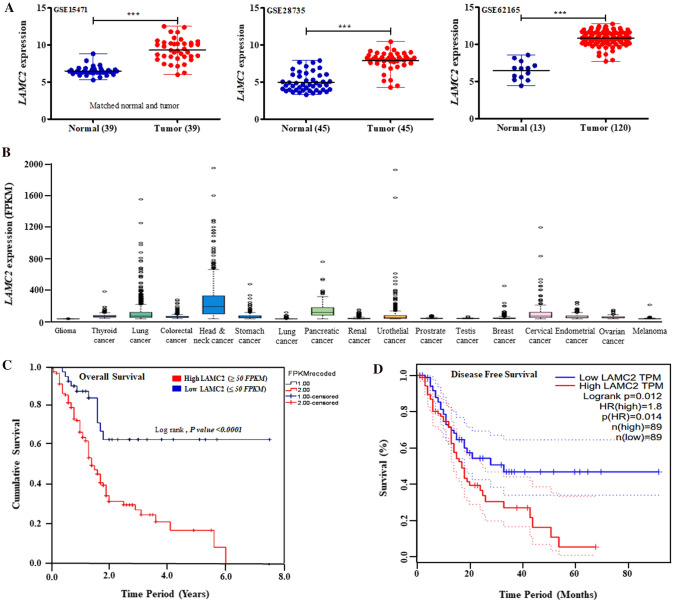
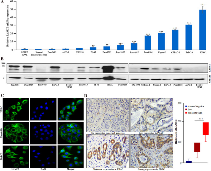
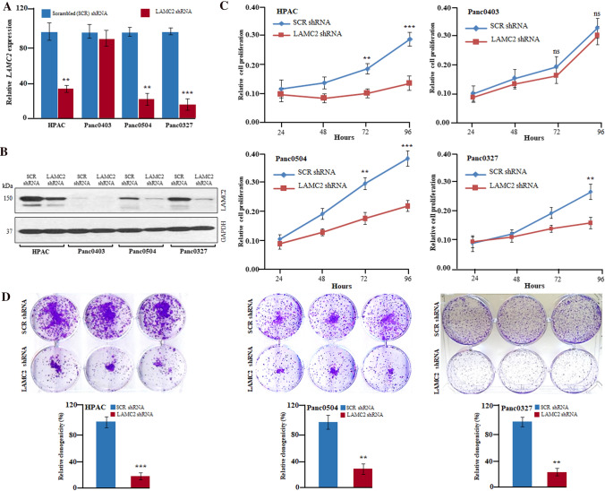
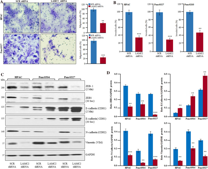
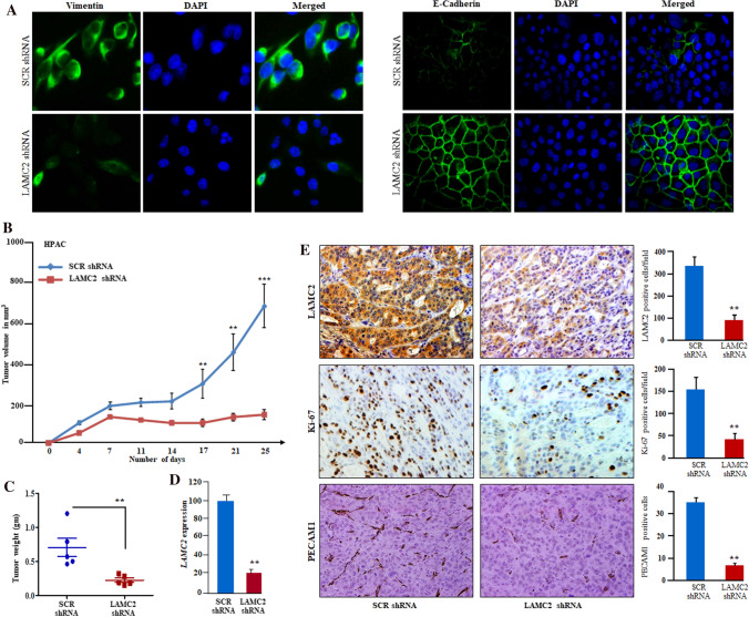
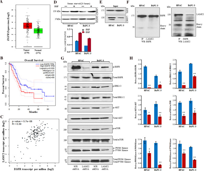
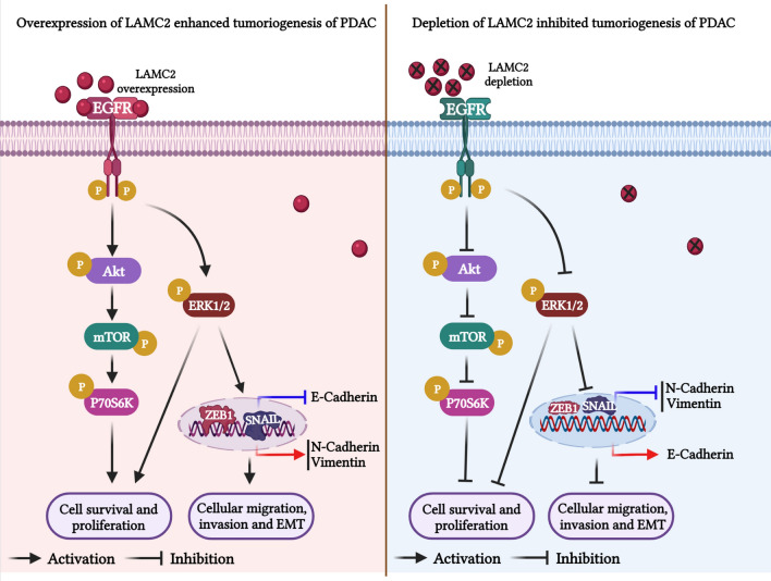
Pancreatic ductal adenocarcinoma (PDAC) is correlated with poor outcomes because of limited therapeutic options. Laminin-5 gamma-2 (LAMC2) plays a critical role in key biological processes. However, the detailed molecular mechanism and potential roles of LAMC2 in PDAC stay unexplored. The present study examines the essential role and molecular mechanisms of LAMC2 in the tumorigenesis of PDAC. Here, we identified that LAMC2 is significantly upregulated in microarray cohorts and TCGA RNA sequencing data of PDAC patients compared to non-cancerous/normal tissues. Patients with higher transcript levels of LAMC2 were correlated with clinical stages; dismal overall, as well as, disease-free survival. Additionally, we confirmed significant upregulation of LAMC2 in a panel of PDAC cell lines and PDAC tumor specimens in contrast to normal pancreatic tissues and cells. Inhibition of LAMC2 significantly decreased cell growth, clonogenic ability, migration and invasion of PDAC cells, and tumor growth in the PDAC xenograft model. Mechanistically, silencing of LAMC2 suppressed expression of ZEB1, SNAIL, N-cadherin (CDH2), vimentin (VIM), and induced E-cadherin (CDH1) expression leading to a reversal of mesenchymal to an epithelial phenotype. Interestingly, co-immunoprecipitation experiments demonstrated LAMC2 interaction with epidermal growth factor receptor (EGFR). Further, stable knockdown of LAMC2 inhibited phosphorylation of EGFR, ERK1/2, AKT, mTOR, and P70S6 kinase signaling cascade in PDAC cells. Altogether, our findings suggest that silencing of LAMC2 inhibited PDAC tumorigenesis and metastasis through repression of epithelial-mesenchymal transition and modulation of EGFR/ERK1/2/AKT/mTOR axis and could be a potential diagnostic, prognostic, and therapeutic target for PDAC.

Keywords: AKT; EGFR; Epithelial-mesenchymal transition; LAMC2; Migration; Pancreatic carcinoma; Xenograft model; mTOR.


Conflict of interest statement

The authors declare that they have no competing interests.

Figures

Fig. 1
Fig. 1
LAMC2 is highly upregulated and associated with worse survival in patients with PDAC. A LAMC2 transcript is overexpressed in PDAC tumor specimens compared to normal or adjacent non-cancerous pancreatic samples in the microarray data (GSE15471, GSE28735, and GSE62125). Statistical significance was calculated using GraphPad Prism (***P-value < 0.0001; two-tailed t-test). B RNA sequencing data analysis displayed higher LAMC2 transcript in PDAC and other human malignancies in TCGA Pan-Cancer datasets. C, D Kaplan–Meier plots analysis displayed elevated levels of LAMC2 transcripts were linked with overall (P-value < 0.0001) and disease-free survival (P-value < 0.012) in the patients with PDAC (TCGA data). Statistical significance was calculated using the Log-rank test
Fig. 2
Fig. 2
Overexpression of LAMC2 transcript and protein in PDAC cell lines and clinical samples. A Quantitative real-time PCR (qPCR) displayed upregulation of LAMC2 transcript in PDAC cells compared to normal human pancreatic tissue and cell line (hTERT-HPNE). Results represent mean ± SD; n = 3. **P-value < 0.001; ***P-value < 0.0001; two-tailed t-test. B Western blot analysis displayed higher LAMC2 protein expression in PDAC cell lines compared to hTERT-HPNE (normal human pancreatic epithelial) cells. C Immunofluorescence experiments demonstrated LAMC2 protein expression in the cytoplasm of PDAC cells. DAPI (4′,6′-diamino-2-phenylindole) was used for nuclear staining. D IHC (immunohistochemistry) data showed strong, moderate, and low staining for LAMC2 in the representative PDAC clinical tissue sections. No/negative staining was noticed in normal pancreatic tissue sections (original magnification, X200). Histoscore analysis displayed significant difference in the staining intensity among low and moderate/high groups. Results represent mean ± SD. ***P-value < 0.0001
Fig. 3
Fig. 3
Inhibition of LAMC2 significantly decreased the growth of PDAC cells in-vitro. A, B. LAMC2 shRNA stable clones of HPAC, Panc0403, Panc0504, and Panc0327 displayed depletion of LAMC2 both at the transcript and protein levels than scramble (SCR) shRNA. GAPDH primers and antibodies were used for normalization and loading controls in qRT-PCR and western blotting experiments, respectively. Experiments were performed thrice in biological triplicates and the results represent mean ± SD. **P-value < 0.001; ***P-value < 0.0001; two-tailed t-test. C. LAMC2 shRNA displayed reduced growth of HPAC, Panc0504, and Panc0327 cells. Results represent mean ± SD; n = 3. **P-value < 0.001; ***P-value < 0.0001; two-tailed t-test. D LAMC2 depletion showed decreased clonogenic growth of HPAC, Panc0504, and Panc0327 cells. Data represent mean ± SD; n = 3. ***P-value < 0.0001;**P-value < 0.001 two-tailed t-test
Fig. 4
Fig. 4
Inhibition of LAMC2 suppressed migration and invasion of PDAC cells through Epithelial-Mesenchymal Transition (EMT). A, B LAMC2 knockdown inhibited migration and invasion of HPAC, Panc0327, and Panc0203 cells. Results represent mean ± SD; n = 3. **P-value < 0.001; ***P-value < 0.0001; two-tailed t-test. C, D Western blotting and densitometry data displayed significantly decreased protein expression of ZEB1, N-cadherin, vimentin, and increased expression of E-cadherin in LAMC2 depleted PDAC cells
Fig. 5
Fig. 5
Inhibition of LAMC2 suppressed the tumor growth in PDAC xenografts. A Immunofluorescence staining of LAMC2 silenced cells displayed expression of E-cadherin on the cell surface and suppressed the expression of vimentin in PDAC cells. DAPI was used for nuclear staining. B Graph displayed decreased tumor volumes and growth in the LAMC2 silenced group compared to the scramble shRNA group (n = 6). C The weight of the tumors in the LAMC2 silenced group was significantly lower (**P-value < 0.001; t-test) than the scramble shRNA group. D qPCR confirmed LAMC2 knockdown in tumor samples. E IHC on tumor section displayed decreased reactivity for LAMC2, Ki-67, and PECAM1 in LAMC2 silenced tumors sections compared to scramble shRNA Tumor sections. Data represent mean ± SD; n = 3. **P-value < 0.001; two-tailed t-test. Original magnification, X200
Fig. 6
Fig. 6
Inhibition of LAMC2 suppressed EGFR/ERK1/2/AKT/mTOR/P70S6K cascade in PDAC. A Upregulation of EGFR transcripts in 179 PDAC samples compared to 171 non-cancerous pancreatic samples in TCGA data. B Higher expression of EGFR displayed inferior overall survival. C Pearson analysis revealed a positive correlation between LAMC2 and EFGR expression. D Epidermal growth factor (100 ng/mL) treatment induced the expression of LAMC2 in HPAC and BxPC-3 cells. E Western blotting experiments displayed robust expression of LAMC2 and EGFR in protein lysates (input) of HPAC and BxPC-3. F LAMC2 antibodies were used for immunoprecipitation in the cell lysates of HPAC and BxPC-3 and immunoprecipitated complexes were immunoblotted with EGFR antibodies. EGFR antibodies were used for immunoprecipitation and immunoprecipitated complexes were subjected to western blotting with LAMC2 antibodies. G HPAC and BxPC-3 cells (either with LAMC2 shRNA or scramble shRNA) were starved for at least 24 h followed by stimulation with EGF (100 ng/mL) for 20 min. Phosphorylation of EGFR, ERK1/2, AKT, mTOR, P70S6K, and P85S6K was demonstrated using western blotting experiments in HPAC and BxPC-3 cells. H Densitometric analysis revealed a significant decrease in phosphorylation of EGFR, ERK1/2, AKT, mTOR, P70S6K, and P85S6K
Fig. 7
Fig. 7
Graphical representation of the molecular mechanism/mode of action of LAMC2 in the tumorigenesis of PDAC. Overexpression of LAMC2 activates EGFR signaling pathway and its downstream signaling by phosphorylating and activating ERK1/2, and AKT/mTOR/P70S6K cascade to enhance the proliferation, survival, migration, invasion, and EMT of PDAC cells. On the other hand, depletion of LAMC2 expression suppressed the phosphorylation of ERK1/2 and AKT/mTOR/P70S6K proteins in HPAC and BxPC-3 cells and reduces the tumorigenesis of PDAC

References

    1. Tempero MA, Malafa MP, Behrman SW, et al. Pancreatic adenocarcinoma, version 2.2014: featured updates to the NCCN guidelines. J Natl Compr Canc Netw. 2014;12:1083–1093. doi: 10.6004/jnccn.2014.0106. - DOI - PubMed
    1. Kleeff J, Korc M, Apte M, et al. Pancreatic cancer. Nat Rev Dis Primers. 2016;2:16022. doi: 10.1038/nrdp.2016.22. - DOI - PubMed
    1. Pandya G, Kirtonia A, Singh A, et al. A comprehensive review of the multifaceted role of the microbiota in human pancreatic carcinoma. Semin Cancer Biol. 2021;26:S1044-579X(21)00157-7. - PubMed
    1. Pandya G, Kirtonia A, Sethi G, Pandey AK, Garg M. The implication of long non-coding RNAs in the diagnosis, pathogenesis and drug resistance of pancreatic ductal adenocarcinoma and their possible therapeutic potential. Biochim Biophys Acta Rev Cancer. 2020;1874:188423. doi: 10.1016/j.bbcan.2020.188423. - DOI - PubMed
    1. Siegel RL, Miller KD, Jemal A. Cancer statistics, 2016. CA Cancer J Clin. 2016;66:7–30. doi: 10.3322/caac.21332. - DOI - PubMed

MeSH terms