. 2022 Jul 15;132(14):e156768.
doi: 10.1172/JCI156768.

Human midbrain dopaminergic neuronal differentiation markers predict cell therapy outcomes in a Parkinson's disease model

Affiliations

Human midbrain dopaminergic neuronal differentiation markers predict cell therapy outcomes in a Parkinson's disease model

Peibo Xu et al. J Clin Invest. .

Abstract

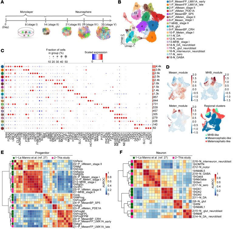
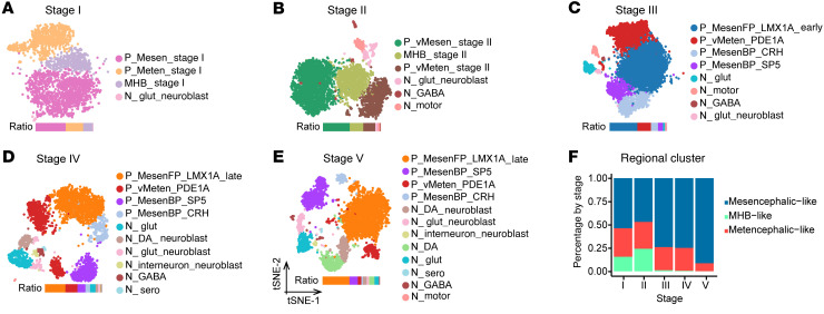
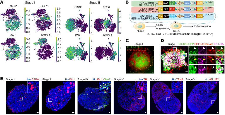
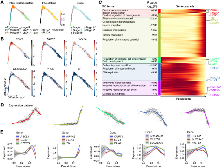
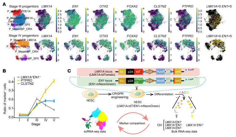
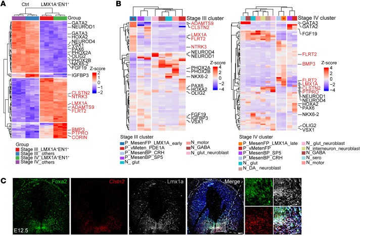
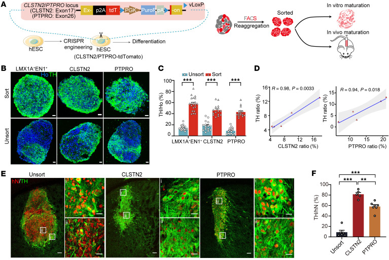
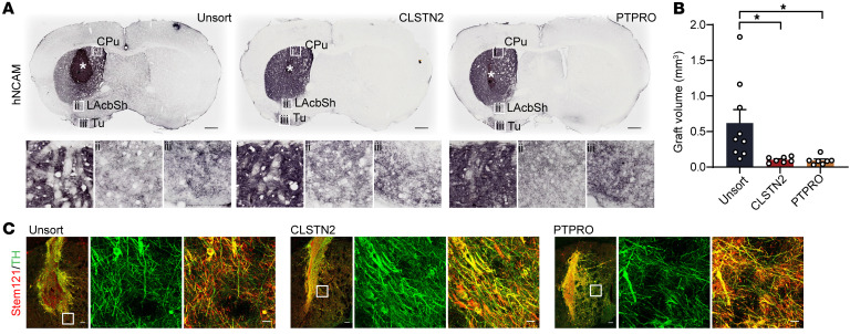
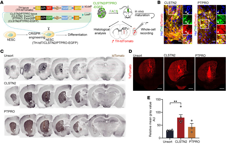
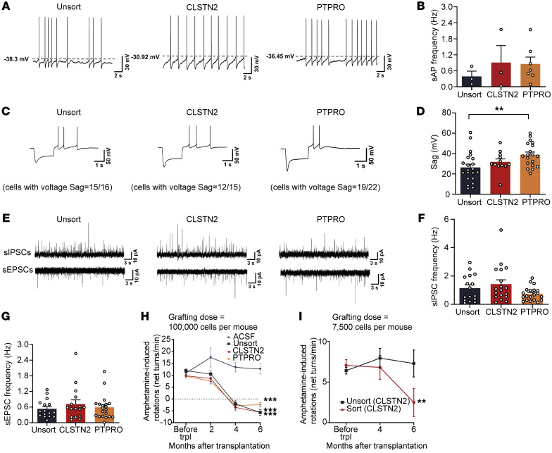
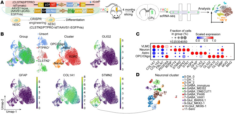
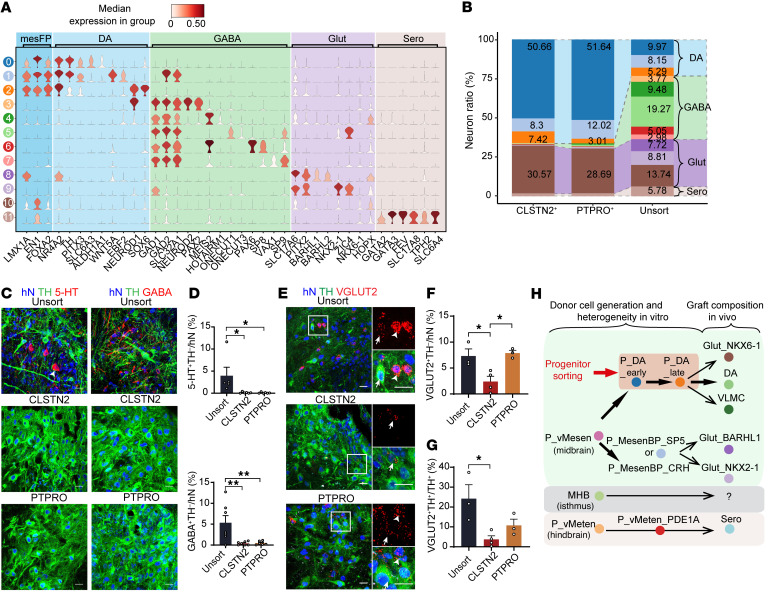
Human pluripotent stem cell-based (hPSC-based) replacement therapy holds great promise for the treatment of Parkinson's disease (PD). However, the heterogeneity of hPSC-derived donor cells and the low yield of midbrain dopaminergic (mDA) neurons after transplantation hinder its broad clinical application. Here, we have characterized the single-cell molecular landscape during mDA neuron differentiation. We found that this process recapitulated the development of multiple but adjacent fetal brain regions including the ventral midbrain, the isthmus, and the ventral hindbrain, resulting in a heterogenous donor cell population. We reconstructed the differentiation trajectory of the mDA lineage and identified calsyntenin 2 (CLSTN2) and protein tyrosine phosphatase receptor type O (PTPRO) as specific surface markers of mDA progenitors, which were predictive of mDA neuron differentiation and could facilitate high enrichment of mDA neurons (up to 80%) following progenitor cell sorting and transplantation. Marker-sorted progenitors exhibited higher therapeutic potency in correcting motor deficits of PD mice. Different marker-sorted grafts had a strikingly consistent cellular composition, in which mDA neurons were enriched, while off-target neuron types were mostly depleted, suggesting stable graft outcomes. Our study provides a better understanding of cellular heterogeneity during mDA neuron differentiation and establishes a strategy to generate highly purified donor cells to achieve stable and predictable therapeutic outcomes, raising the prospect of hPSC-based PD cell replacement therapies.

Keywords: Neuroscience; Parkinson disease; Stem cell transplantation; Stem cells.


Figures

Figure 1
Figure 1. Time-course single-cell RNA-Seq reveals cellular heterogeneity during differentiation of mDA neurons from hPSCs.
(A) Schematics of in vitro cell differentiation process for scRNA-Seq. (B) Visualization of clustering results of merged data sets from all stages using uniform manifold approximation and projection (UMAP). (C) Dot plot showing classical markers of floor plate and representative markers (column) for each cell type (row). Mean gene expression has been scaled between 0 and 1. Horizontal bars denote the number of cells in each cluster. Cell-type labels are used as UMAP clusters in B. The dot color scale represents average expression levels, and dot size represents the fraction of cells in a group. (D) Regional gene module expression and regional annotation of time-course scRNA-Seq data. See Supplemental Methods for the module gene lists used to calculate the gene expression scores. (E and F) Heatmaps of area under the receiver operating characteristic (AUROC) scores between progenitor (E) and neuron (F) clusters in this study and data from a public data set (27). P, progenitor; N, neuron; vMesen, ventral mesencephalic; vMeten, ventral metencephalic; MesenFP, mesencephalic floor plate; MesenBP, mesencephalic basal plate.
Figure 2
Figure 2. Cell-type composition at each stage of mDA neuron differentiation.
(A–E) Visualization of clustering results for each stage using t-distributed stochastic neighbor embedding (t-SNE). (F) Change in the percentage of regional clusters by stage. The MHB-like cluster mainly emerged at the early patterning stage (stages I and II).
Figure 3
Figure 3. The process of mDA neuron differentiation recapitulates the development of adjacent fetal brain regions including the ventral midbrain, the isthmus, and the ventral hindbrain.
(A) Stage I or II scRNA-Seq clusters of cells showing expression of OTX2, FGF8, EN1, and HOXA2 genes. (B) OTX2/FGF8/EN1 reporter cell line diagram. (C) Typical colony of stage I EGFP/tdT cells by live imaging. (D) Typical colony of stage I EGFP/tdT cells by immunostaining for EGFP/tdT/HA-tag. The white arrowhead, yellow arrowhead, and white arrow indicate an FGF8-tdT+ cell, FGF8-tdT+EN1-HA+ cell, and OTX2-EGFP+EN1-HA+ cell, respectively. Scale bars: 50 μm (C) and 25 μm (D). Original magnification, ×20 (enlarged insets in D). (E) Typical neurospheres immunostained for neuronal markers at distinct stages. VGLUT2 was validated by RNA-FISH (RNAScope) and the others by antibodies. Scale bars: 25 μm.
Figure 4
Figure 4. Reconstruction of single-cell trajectory of mDA neuron differentiation reveals a dynamic and characteristic lineage-specific transcriptional profile.
(A) Visualization using diffusion map embeddings by mDA-related clusters, pseudotime, and stage. (B) Typical marker expression on a diffusion map. (C) Enriched GO terms for each gene cluster (left) and gene expression cascade (right) during mDA differentiation. Heatmap shows selected gene expression along pseudotime. Expression is displayed as the mean expression of groups of 5 cells and was smoothed using a spline curve and scaled to the maximum observed expression (low expression in yellow, high expression in red). The colored label along the left side of the heatmap identifies the gene cluster. (D) Mean expression profiles for each gene cluster. The colors of the spline curves correspond to the gene cluster colors in C. (E) Expression of selected genes for each gene cluster. The curve represents the mean expression of the gene, and the standard error of the mean is shown as a gray band.
Figure 5
Figure 5. CLSTN2 and PTPRO are identified as specific surface markers for early and late mDA progenitors, respectively.
(A) Progenitor clusters; expression of mDA progenitor marker genes (LMX1A, EN1, OTX2, and FOXA2) and of 2 identified surface marker genes (CLSTN2, PTPRO); and annotated LMX1A+EN1+ cells (LMX1A unique molecular identifier [UMI] counts >0 and EN1 UMI counts >0) on UMAP embeddings of stage III and stage IV progenitors. (B) Selected marker-positive cell ratio for a random set of cells (10% of cells from each stage). Data represent the mean ± SD. See Supplemental Methods for details. (C) Dual-reporter cell line diagram and schematics of joint analysis of bulk RNA-Seq and scRNA-Seq data. Ctrl, control.
Figure 6
Figure 6. Identified surface markers are coexpressed with classical mDA progenitor markers in vitro and in developing mouse ventral midbrain.
(A) Heatmap showing scaled expression of 4 groups of top 40 DEGs (stage III LMX1A+EN1+ and control; stage IV LMX1A+EN1+ and control). Control cells were collected from LMX1AEN1, LMX1A+EN1, and LMX1AEN1+. DEGs in LMX1A+EN1+ cells are shown in red, and DEGs in control cells are shown in black. (B) Heatmap showing scaled expression of same DEGs from A projected onto stage III (left) and stage IV (right) scRNA-Seq clusters. Marker genes are shown in the same color as in A. (C) RNA-FISH of Clstn2 following IHC by colabeling Foxa2 and Lmx1a in E12.5 mouse mesencephalon. The zoomed views indicate magnified images of Foxa2+Lmx1a+Clstn2+ progenitors. Scale bars: 100 μm. Original magnification, ×20 (higher-magnification images).
Figure 7
Figure 7. CLSTN2 and PTPRO are predictive of mDA neuron differentiation and can give rise to highly enriched mDA neurons after progenitor sorting and transplantation.
(A) Diagram of surface marker reporter cell lines and experimental schematics for in vitro and in vivo maturation. (B and C) Neurospheres matured in vitro and (B) immunostained for TH and (C) statistical analysis (n = 3 batches with 5 neurospheres per batch). Scale bars: 25 μm. ***P < 0.001, by multiple unpaired t test with Holm-Šidák correction. (D) Correlation between the surface marker progenitor ratio and the TH+ neuron ratio. See Figure 9A for a diagram of the cell lines used. (E) Unsorted progenitor-, CLSTN2+ progenitor–, and PTPRO+ progenitor–derived grafts immunostained for human nuclei (hN) and TH. Scale bars: 100 μm and 20 μm (for the enlarged insets [i] and [ii], which represent the edge and center area of the graft, respectively). (F) Quantification of the TH+ neurons ratio in grafts. n = 6 (unsorted), n = 5 (CLSTN2), and n = 7 (PTPRO). **P < 0.01 and ***P < 0.001, by 1-way ANOVA followed by Tukey’s multiple-comparison test. Sort, sorted; unsort, unsorted.
Figure 8
Figure 8. CLSTN2- or PTPRO-enriched progenitors reinnervate the host striatum and give rise to smaller grafts.
(A) Immunostaining for hNCAM in grafted neurons showed hNCAM+ fiber distribution and extension into the dorsal striatum (caudate putamen [CPu], inset box i) and the ventral striatum (lateral nucleus accumbens shell [LAcbSh], inset box ii; olfactory tubercle [Tu], inset box iii). White asterisk indicates the graft site. Scale bars: 500 μm. (B) Graft volumes were estimated by hN staining at 6 months. n = 9 (unsorted), n = 7 (CLSTN2), n = 8 (PTPRO). *P < 0.05, by 1-way ANOVA followed by Tukey’s multiple-comparison test. (C) Grafts were colabeled for human-specific fiber STEM121 and TH. Scale bars: 100 μm and 20 μm (insets, representing zoomed views of extended graft fiber).
Figure 9
Figure 9. CLSTN2- or PTPRO-enriched progenitors give rise to denser DA innervations after transplantation.
(A) Schematics for TH-specific histological evaluation and electrophysiological recording in surface marker–derived grafts. (B) Immunostaining for tdT in TH+ neurons in CLSTN2- and PTPRO-derived grafts. Boxed areas are magnified on the right. White arrows indicate neurons coexpressing tdT and TH. Scale bars: 20 μm. Original magnification, ×60 (enlarged insets). (C) Serial coronal sections of grafts immunostained for tdT. White asterisk indicates the graft site. Scale bars: 500 μm. (D) Typical IHC images with tdT labeling (representing TH) in grafts. Scale bars: 500 μm. (E) Quantification of the mean gray value of tdT pixels from 4 random areas within the host striatum (see Supplemental Methods for details). **P < 0.01, by 1-way ANOVA followed by Tukey’s multiple-comparison test.
Figure 10
Figure 10. CLSTN2- or PTPRO-enriched progenitors integrate into host circuits and exhibit a higher therapeutic potency.
(AD) Typical traces of whole-cell patch-clamp recording of sAPs (A) and sAP frequency (B), hyperpolarizing current injection showing voltage sag (C), and voltage sag measurements (D) from grafted mDA neurons 5 months after transplantation. Recorded cell numbers: n = 24 (unsorted), n = 15 (CLSTN2), n = 22 (PTPRO). **P < 0.01, by 1-way ANOVA followed by Tukey’s multiple-comparison test. (E) Typical traces of sIPSCs (top) and sEPSCs (bottom) in grafted human mDA neurons 5 months after transplantation. (F and G) Frequencies of sIPSCs (F) and sEPSCs (G). Number of mice: n = 4 (unsorted), n = 3 (CLSTN2), n = 4 (PTPRO). Recorded cell numbers for sEPSCs: n = 16 (unsorted), n = 16 (CLSTN2), n = 20 (PTPRO). Recorded cell numbers for sIPSCs: n = 16 (unsorted), n = 18 (CLSTN2), n = 20 (PTPRO). (H and I) Amphetamine-induced rotation behavior changes in PD mice over a 6-month post-transplantation period. The grafting dose per mouse was 100,000 cells (H). n = 5 (aCSF), n = 9 (unsorted), n = 11 (CLSTN2), n = 9 (PTPRO). (I) The grafting dose per mouse was 7500 cells. The H9-CLSTN2-P2A-tdT cell line was used. n = 4 (unsorted), n = 3 (sorted). The tdT ratio for the unsorted group was approximately 29%. **P < 0.01 and ***P < 0.001, by 2-way ANOVA with Dunnett’s multiple-comparison test, compared with the ACSF group (H) or with the unsorted group (I). trpl, transplantation.
Figure 11
Figure 11. scRNA-Seq reveals the cellular composition of grafts.
(A) Schematics for scRNA-Seq of grafts. (B) Clustering recovered 4 major cell types in grafts and their corresponding typical gene expression. (C) Dot plot showing markers of the cell types in grafts. (D) Further clustering of neurons.
Figure 12
Figure 12. Grafts from CLSTN2- or PTPRO-enriched progenitors contain enriched mDA neurons with most off-target neuron types depleted.
(A) Violin plot of representative markers for all graft neuronal clusters. (B) Neuronal subtype ratio for each graft group (unsorted, CLSTN2, and PTPRO). (C and D) Typical images of grafts immunostained for 5-HT (C, left) or GABA (C, right) and the mDA marker TH. Quantification of 5-HT+ (D, top) and GABA+ neuron ratios (D, bottom) in grafts for each group. *P < 0.05 and **P < 0.01, by 1-way ANOVA followed by Tukey’s multiple-comparison test. (EG) Representative images of grafts immunostained for VGLUT2 (RNAScope) and TH (E). Arrowheads and arrows indicate VGLUT2+TH neurons and TH+ neurons with weak VGLUT2 expression, respectively. Quantification of VGLUT2+TH/hN ratio (F) and the VGLUT2+TH+/TH+ ratio (G). Scale bars: 20 μm. *P < 0.05, by 1-way ANOVA followed by Tukey’s multiple-comparison test. (H) Proposed model of how heterogenous donor cells generated in vitro result in grafts with diverse neuronal composition in vivo. Graft outcomes can be improved and predicted after mDA progenitor sorting via specific markers.

Comment in

References

    1. Parmar M, et al. The future of stem cell therapies for Parkinson disease. Nat Rev Neurosci. 2020;21(2):103–115. doi: 10.1038/s41583-019-0257-7. - DOI - PubMed
    1. Barker RA, et al. Human trials of stem cell-derived dopamine neurons for Parkinson’s Disease: dawn of a new era. Cell Stem Cell. 2017;21(5):569–573. doi: 10.1016/j.stem.2017.09.014. - DOI - PubMed
    1. Yamanaka S. Pluripotent stem cell-based cell therapy-promise and challenges. Cell Stem Cell. 2020;27(4):523–531. doi: 10.1016/j.stem.2020.09.014. - DOI - PubMed
    1. Kim TW, et al. Pluripotent stem cell therapies for parkinson disease: present challenges and future opportunities. Front Cell Dev Biol. 2020;8:729. doi: 10.3389/fcell.2020.00729. - DOI - PMC - PubMed
    1. Kirkeby A, et al. Generation of regionally specified neural progenitors and functional neurons from human embryonic stem cells under defined conditions. Cell Rep. 2012;1(6):703–714. doi: 10.1016/j.celrep.2012.04.009. - DOI - PubMed

Publication types