

3T-MRI-based age, sex and site-specific markers of musculoskeletal health in healthy children and young adults
- PMID: 35700237
- PMCID: PMC9346338
- DOI: 10.1530/EC-22-0034
3T-MRI-based age, sex and site-specific markers of musculoskeletal health in healthy children and young adults
Abstract
Objective: The aim of this study is to investigate the role of 3T-MRI in assessing musculoskeletal health in children and young people.
Design: Bone, muscle and bone marrow imaging was performed in 161 healthy participants with a median age of 15.0 years (range, 8.0, 30.0).
Methods: Detailed assessment of bone microarchitecture (constructive interference in the steady state (CISS) sequence, voxel size 0.2 × 0.2 × 0.4 mm3), bone geometry (T1-weighted turbo spin echo (TSE) sequence, voxel size 0.4 × 0.4 × 2 mm3) and bone marrow (1H-MRS, point resolved spectroscopy sequence (PRESS) (single voxel size 20 × 20 × 20 mm3) size and muscle adiposity (Dixon, voxel size 1.1 × 1.1 × 2 mm3).
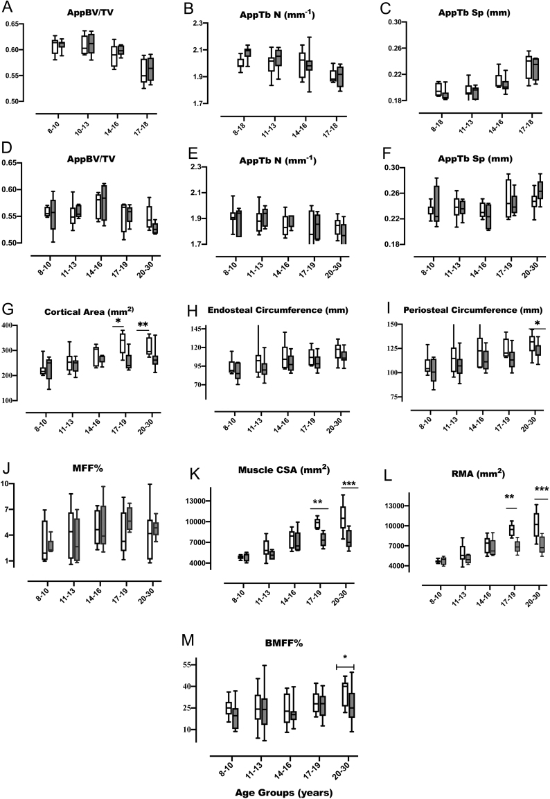
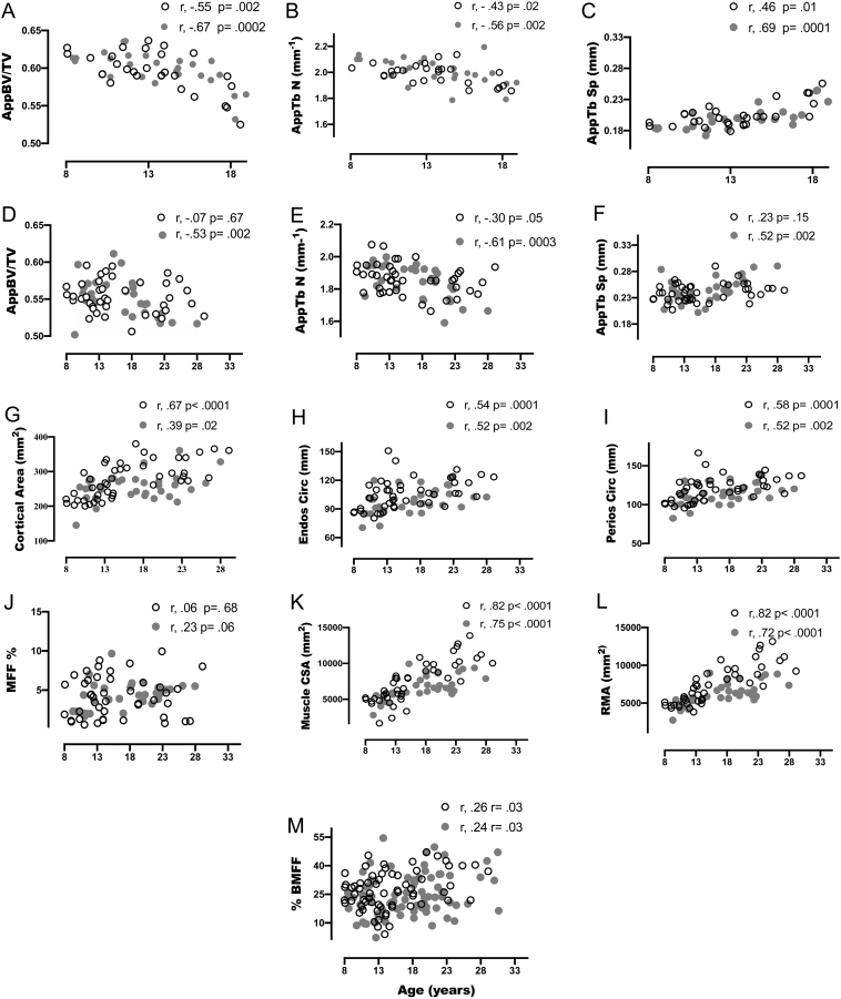
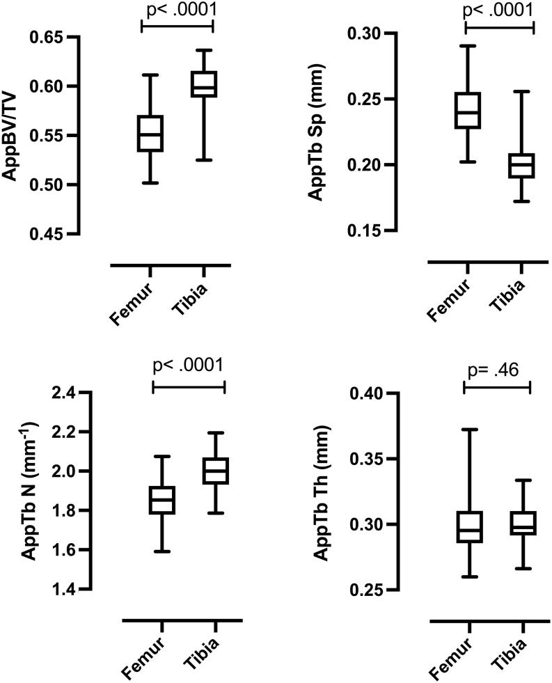
Results: There was an inverse association of apparent bone volume/total volume (appBV/TV) with age (r = -0.5, P < 0.0005). Cortical area, endosteal and periosteal circumferences and muscle cross-sectional area showed a positive association to age (r > 0.49, P < 0.0001). In those over 17 years of age, these parameters were also higher in males than females (P < 0.05). This sex difference was also evident for appBV/TV and bone marrow adiposity (BMA) in the older participants (P < 0.05). AppBV/TV showed a negative correlation with BMA (r = -0.22, P = 0.01) which also showed an association with muscle adiposity (r = 0.24, P = 0.04). Cortical geometric parameters were highly correlated with muscle area (r > 0.57, P < 0.01).
Conclusions: In addition to providing deep insight into the normal relationships between bone, fat and muscle in young people, these novel data emphasize the role of MRI as a non-invasive method for performing a comprehensive and integrated assessment of musculoskeletal health in the growing skeleton.
Keywords: MRI; adiposity; bone; marrow; muscle.
Figures

Similar articles
-
MRI-based abnormalities in young adults at risk of adverse bone health due to childhood-onset metabolic & endocrine conditions.Clin Endocrinol (Oxf). 2014 Jun;80(6):811-7. doi: 10.1111/cen.12367. Epub 2013 Dec 12. Clin Endocrinol (Oxf). 2014. PMID: 24245820
-
Deficits in Trabecular Bone Microarchitecture in Young Women With Type 1 Diabetes Mellitus.J Bone Miner Res. 2015 Aug;30(8):1386-93. doi: 10.1002/jbmr.2465. J Bone Miner Res. 2015. PMID: 25627460 Clinical Trial.
-
Muscle deficits with normal bone microarchitecture and geometry in young adults with well-controlled childhood-onset Crohn's disease.Eur J Gastroenterol Hepatol. 2020 Dec;32(12):1497-1506. doi: 10.1097/MEG.0000000000001838. Eur J Gastroenterol Hepatol. 2020. PMID: 32675776
-
Visceral adiposity and inflammatory bowel disease.Int J Colorectal Dis. 2021 Nov;36(11):2305-2319. doi: 10.1007/s00384-021-03968-w. Epub 2021 Jun 9. Int J Colorectal Dis. 2021. PMID: 34104989 Review.
-
Reporting Guidelines, Review of Methodological Standards, and Challenges Toward Harmonization in Bone Marrow Adiposity Research. Report of the Methodologies Working Group of the International Bone Marrow Adiposity Society.Front Endocrinol (Lausanne). 2020 Feb 28;11:65. doi: 10.3389/fendo.2020.00065. eCollection 2020. Front Endocrinol (Lausanne). 2020. PMID: 32180758 Free PMC article. Review.
Cited by
-
The influence of age and sex on speed-strength performance in children between 10 and 14 years of age.Front Physiol. 2023 Feb 21;14:1092874. doi: 10.3389/fphys.2023.1092874. eCollection 2023. Front Physiol. 2023. PMID: 36895629 Free PMC article.
References
Grants and funding
LinkOut - more resources
Full Text Sources
