

Site-specific glycosylation of SARS-CoV-2: Big challenges in mass spectrometry analysis
- PMID: 35700310
- PMCID: PMC9349404
- DOI: 10.1002/pmic.202100322
Site-specific glycosylation of SARS-CoV-2: Big challenges in mass spectrometry analysis
Abstract
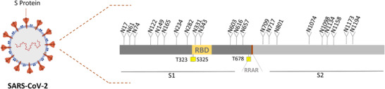
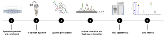
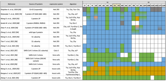
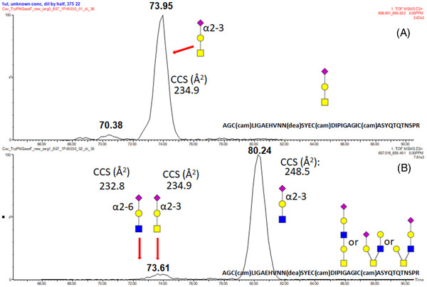
Glycosylation of viral proteins is required for the progeny formation and infectivity of virtually all viruses. It is increasingly clear that distinct glycans also play pivotal roles in the virus's ability to shield and evade the host's immune system. Recently, there has been a great advancement in structural identification and quantitation of viral glycosylation, especially spike proteins. Given the ongoing pandemic and the high demand for structure analysis of SARS-CoV-2 densely glycosylated spike protein, mass spectrometry methodologies have been employed to accurately determine glycosylation patterns. There are still many challenges in the determination of site-specific glycosylation of SARS-CoV-2 viral spike protein. This is compounded by some conflicting results regarding glycan site occupancy and glycan structural characterization. These are probably due to differences in the expression systems, form of expressed spike glycoprotein, MS methodologies, and analysis software. In this review, we recap the glycosylation of spike protein and compare among various studies. Also, we describe the most recent advancements in glycosylation analysis in greater detail and we explain some misinterpretation of previously observed data in recent publications. Our study provides a comprehensive view of the spike protein glycosylation and highlights the importance of consistent glycosylation determination.
Keywords: N-glycosylation; O-glycosylation; SARS-CoV-2 glycoprotein; glycoproteomics.
© 2022 The Authors. Proteomics published by Wiley-VCH GmbH.
Conflict of interest statement
The authors declare no conflict of interest.
Figures

Similar articles
-
Analysis of the N-glycosylation profiles of the spike proteins from the Alpha, Beta, Gamma, and Delta variants of SARS-CoV-2.Anal Bioanal Chem. 2023 Aug;415(19):4779-4793. doi: 10.1007/s00216-023-04771-y. Epub 2023 Jun 24. Anal Bioanal Chem. 2023. PMID: 37354227 Free PMC article.
-
Data-independent acquisition mass spectrometry for site-specific glycoproteomics characterization of SARS-CoV-2 spike protein.Anal Bioanal Chem. 2021 Dec;413(29):7305-7318. doi: 10.1007/s00216-021-03643-7. Epub 2021 Oct 12. Anal Bioanal Chem. 2021. PMID: 34635934 Free PMC article.
-
Comparison of N- and O-Glycosylation on Spike Glycoprotein 1 of SARS-CoV-1 and MERS-CoV.J Proteome Res. 2025 May 2;24(5):2256-2265. doi: 10.1021/acs.jproteome.4c00716. Epub 2025 Apr 7. J Proteome Res. 2025. PMID: 40193531
-
Glycosylation of SARS-CoV-2: structural and functional insights.Anal Bioanal Chem. 2021 Dec;413(29):7179-7193. doi: 10.1007/s00216-021-03499-x. Epub 2021 Jul 7. Anal Bioanal Chem. 2021. PMID: 34235568 Free PMC article. Review.
-
Site-Specific O-glycosylation of SARS-CoV-2 Spike Protein and Its Impact on Immune and Autoimmune Responses.Cells. 2024 Jan 5;13(2):107. doi: 10.3390/cells13020107. Cells. 2024. PMID: 38247799 Free PMC article. Review.
Cited by
-
Variations in O-Glycosylation Patterns Influence Viral Pathogenicity, Infectivity, and Transmissibility in SARS-CoV-2 Variants.Biomolecules. 2023 Sep 29;13(10):1467. doi: 10.3390/biom13101467. Biomolecules. 2023. PMID: 37892149 Free PMC article.
-
Screening and Characterization of Shark-Derived VNARs against SARS-CoV-2 Spike RBD Protein.Int J Mol Sci. 2022 Sep 18;23(18):10904. doi: 10.3390/ijms231810904. Int J Mol Sci. 2022. PMID: 36142819 Free PMC article.
-
Water-glycan interactions drive the SARS-CoV-2 spike dynamics: insights into glycan-gate control and camouflage mechanisms.Chem Sci. 2024 Aug 23;15(35):14177-87. doi: 10.1039/d4sc04364b. Online ahead of print. Chem Sci. 2024. PMID: 39220162 Free PMC article.
-
Analysis of carbohydrates and glycoconjugates by matrix-assisted laser desorption/ionization mass spectrometry: An update for 2021-2022.Mass Spectrom Rev. 2025 May-Jun;44(3):213-453. doi: 10.1002/mas.21873. Epub 2024 Jun 24. Mass Spectrom Rev. 2025. PMID: 38925550 Free PMC article. Review.
-
Inclusion of deuterated glycopeptides provides increased sequence coverage in hydrogen/deuterium exchange mass spectrometry analysis of SARS-CoV-2 spike glycoprotein.Rapid Commun Mass Spectrom. 2024 Mar 15;38(5):e9690. doi: 10.1002/rcm.9690. Rapid Commun Mass Spectrom. 2024. PMID: 38355883 Free PMC article.
References
-
- Braun, L. , Brenier‐Pinchart, M.‐P. , Yogavel, M. , Curt‐Varesano, A. , Curt‐Bertini, R.‐L. , Hussain, T. , Kieffer‐Jaquinod, S. , Coute, Y. , Pelloux, H. , Tardieux, I. , Sharma, A. , Belrhali, H. , Bougdour, A. , & Hakimi, M.‐A. (2013). A Toxoplasma dense granule protein, GRA24, modulates the early immune response to infection by promoting a direct and sustained host p38 MAPK activation. Journal of Experimental Medicine, 210(10), 2071–2086. 10.1084/jem.20130103 - DOI - PMC - PubMed
-
- Duyen, H. T. L. , Ngoc, T. V. , Ha, D. T. , Hang, V. T. T. , Kieu, N. T. T. , Young, P. R. , Farrar, J. J. , Simmons, C. P. , Wolbers, M. , & Wills, B. A. (2011). Kinetics of plasma viremia and soluble nonstructural protein 1 concentrations in dengue: Differential effects according to serotype and immune status. Journal of Infectious Diseases, 203(9), 1292–1300. 10.1093/infdis/jir014 - DOI - PMC - PubMed
Publication types
MeSH terms
Substances
LinkOut - more resources
Full Text Sources
Medical
Miscellaneous
