. 2022 Jun 21;119(25):e2122477119.
doi: 10.1073/pnas.2122477119. Epub 2022 Jun 14.

Single-dose ethanol intoxication causes acute and lasting neuronal changes in the brain

Affiliations

Single-dose ethanol intoxication causes acute and lasting neuronal changes in the brain

Johannes Knabbe et al. Proc Natl Acad Sci U S A. .

Abstract

Alcohol intoxication at early ages is a risk factor for the development of addictive behavior. To uncover neuronal molecular correlates of acute ethanol intoxication, we used stable-isotope-labeled mice combined with quantitative mass spectrometry to screen more than 2,000 hippocampal proteins, of which 72 changed synaptic abundance up to twofold after ethanol exposure. Among those were mitochondrial proteins and proteins important for neuronal morphology, including MAP6 and ankyrin-G. Based on these candidate proteins, we found acute and lasting molecular, cellular, and behavioral changes following a single intoxication in alcohol-naïve mice. Immunofluorescence analysis revealed a shortening of axon initial segments. Longitudinal two-photon in vivo imaging showed increased synaptic dynamics and mitochondrial trafficking in axons. Knockdown of mitochondrial trafficking in dopaminergic neurons abolished conditioned alcohol preference in Drosophila flies. This study introduces mitochondrial trafficking as a process implicated in reward learning and highlights the potential of high-resolution proteomics to identify cellular mechanisms relevant for addictive behavior.

Keywords: Drosophila; addiction; ethanol; plasticity; two-photon microscopy.


Conflict of interest statement

The authors declare no competing interest.

Figures
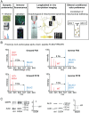
Fig. 1.
Fig. 1.
Study design and quantitative MS of synaptic proteomes. (A) Study design. (B) Raw MS spectra of WT and SILAC peptides. Reproducible and significant ethanol-dependent changes in peptide abundance of a peptide (FLSDVYPDGFK) unique to PCCA. In all four experiments, ethanol exposure roughly doubled the peak height compared to untreated controls. (C) WB analysis of two candidate proteins. Actin was used as a loading control as MS results showed that the actin protein did not change between ethanol (EtOH) and untreated control (ctrl) conditions. Numbers indicate molecular weight markers. Graphs show quantifications (black dots) of two independent WB experiments for each candidate protein. The ratio of candidate protein/actin under untreated conditions was normalized to 1 for comparison with the ratio of candidate protein/actin after ethanol stimulation.
Fig. 2.
Fig. 2.
Immunofluorescence detection of ethanol-dependent synaptic protein dynamics in vivo. (A–D) Images for ankyrin-G analysis. (E–H) Images for MAP6 analysis. (I–L) Images for PCCA analysis. (B) Ankyrin-G stain. (F) MAP6 stain. (J) PCCA stain. (A, E, and I) β-actin stain. (C, G, and K) Synapsin stain. Merged images (D, H, and L). Insets: high magnification of synaptic labels (green: β-actin, red: ankyrin-G/MAP6/PCCA, blue: synapsin). (M) Quantification of ankyrin-G/synapsin ratios over time, at 1, 2, 3, 4, 6, or 24 h after 3.5 g/kg ethanol i.p. injection. Each sample point corresponds to a single image as shown in this figure; each time point was based on one mouse. The y axis shows the synaptic ratio of [fluorescence intensity candidate protein]/[fluorescence intensity synapsin]. (N) Quantification of MAP6/synapsin ratios over time. (O) Quantification of PCCA/synapsin ratios over time (Scale bars, A–L, 20 µm; insets, 1 µm).
Fig. 3.
Fig. 3.
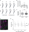
Neuronal in vivo ethanol-dependent morphological plasticity. (A) Longitudinal in vivo two-photon imaging of the same dendritic stretches in Thy1-GFP mice identified with stable (blue) and unstable spines (red). (B) Quantification of in vivo spine turnover from longitudinal imaging data (P < 0.05; two-way repeated measures ANOVA with Bonferroni’s multiple comparisons test; n = four mice; error bars, SD). (C) Ethanol intoxication in mice did not induce changes in cortical spine density after 4 to 6 h (six mice for each condition; all mice received pyrazole), n.s., nonsignificant. (D) Immunofluorescence detection of the AIS in layer II/III pyramidal neurons of S1 (green: βIV−spectrin; purple: NeuN) (Scale bar, 10 µm). (E) Change in AIS length over time after a single ethanol i.p. injection (3.5 g/kg ethanol); approximately 100 AIS structures were measured for each data point (P = 0.021; n = seven mice; error bars, SD). *P < 0.05.
Fig. 4.
Fig. 4.
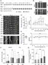
In vivo imaging of mitochondria and presynaptic boutons reveals ethanol-dependent effects. (A) For each mouse, the experiment consisted of four imaging sessions. Baseline imaging (unfilled boxes) was acquired each day. Saline (day 1, light gray arrow) and ethanol (day 3, dark gray arrow) were administered i.p. followed by postinjection imaging (filled boxes). On days 2 and 4, 24 h postinjection images were recorded. Time lapses (10 min mitochondria; 5 min DCVs) were acquired every 30 min. (B) Example of a kymograph before ethanol injection (Top); only one mitochondrion was mobile. Kymograph of the same axonal stretch 180 min after ethanol injection (Bottom). (C) Top, axonal stretch at different time points (y axis, seconds). Bottom, corresponding kymograph. (D) Left, overall mitochondrial mobility. Right, time course of mitochondrial mobility under basal conditions (light gray) and after ethanol injection (dark gray) (n = 11 focal planes from four mice; mean ± SD). (E) Left, overall DCV mobility. Right, time course of DCV mobility under basal conditions (light gray) and after ethanol injection (dark gray) (n = 3 mice; mean ± SD). (F) Bouton turnover at different time points. (G) Loss of unoccupied presynaptic boutons was significantly increased after 4 h and 24 h following ethanol injection (n = 40 axons from four mice, mean ± SD). Saline: light gray, ethanol: dark gray for all graphs. Z = Z-stack, T = time lapse. *P < 0.05, **P < 0.01, ***P < 0.001, ****P <0.0001, ns, P > 0.05, nonsignificant.
Fig. 5.
Fig. 5.
The function of ethanol as positive reinforcer in dopaminergic neurons depends on Miro and Milton. (A) The expression of UAS-dmiro-RNAiTRiPJF02775 under the control of the TH-Gal4 driver resulted in the loss of CPI (conditioned odor preference index). The mean of the CPI was for TH-Gal4/+: 0.22 ± 0.07; UAS-dmiro-RNAi/+: 0.21 ± 0.04 and for the experimental group −0.05 ± 0.07. (B) The expression of the UAS-milton-RNAiGD8116 and UAS-milton-RNAiTRiPJF03022 under the control of the TH-Gal4 driver resulted in loss of CPI. The mean of the CPI was for TH-Gal4/+: 0.37 ± 0.08, UAS-milton-RNAiGD8116/+: 0.31 ± 0.04 and for the experimental group 0.05 ± 0.06 and TH-Gal4/+: 0.32 ± 0.07; for UAS-milton-RNAiTRiPJF03022/+: 0.19 ± 0.03 and for the experimental group −0.01 ± 0.06. The letter “a” indicates significant differences from random choice as determined with the 1-sample sign test. Differences between groups were determined using ANOVA posthoc Tukey–Kramer HSD (honestly significant difference) (Error bars, SEM).

Comment in

References

    1. DeWit D. J., Adlaf E. M., Offord D. R., Ogborne A. C., Age at first alcohol use: A risk factor for the development of alcohol disorders. Am. J. Psychiatry 157, 745–750 (2000). - PubMed
    1. Morean M. E., et al. , First drink to first drunk: Age of onset and delay to intoxication are associated with adolescent alcohol use and binge drinking. Alcohol. Clin. Exp. Res. 38, 2615–2621 (2014). - PMC - PubMed
    1. Whelan R., et al. ; IMAGEN Consortium, Neuropsychosocial profiles of current and future adolescent alcohol misusers. Nature 512, 185–189 (2014). - PMC - PubMed
    1. Henry K. L., et al. , Age of onset of first alcohol intoxication and subsequent alcohol use among urban American Indian adolescents. Psychol. Addict. Behav. 25, 48–56 (2011). - PMC - PubMed
    1. Hingson R., Heeren T., Zakocs R., Winter M., Wechsler H., Age of first intoxication, heavy drinking, driving after drinking and risk of unintentional injury among U.S. college students. J. Stud. Alcohol 64, 23–31 (2003). - PubMed

Publication types

MeSH terms

LinkOut - more resources