

An ecological approach to identify distinct neural correlates of disinhibition in frontotemporal dementia
- PMID: 35700600
- PMCID: PMC9194654
- DOI: 10.1016/j.nicl.2022.103079
An ecological approach to identify distinct neural correlates of disinhibition in frontotemporal dementia
Abstract
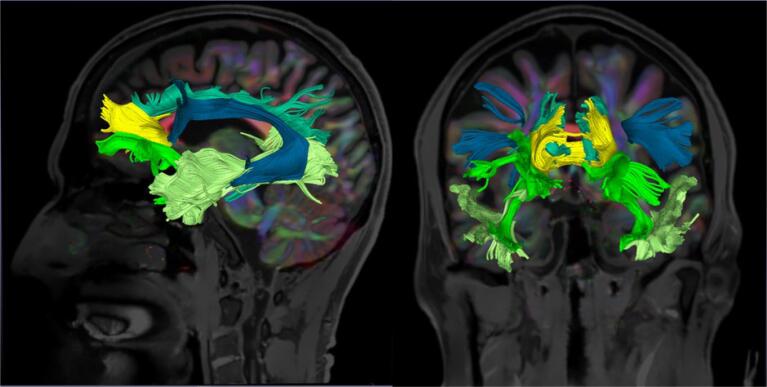
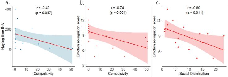
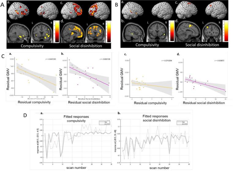
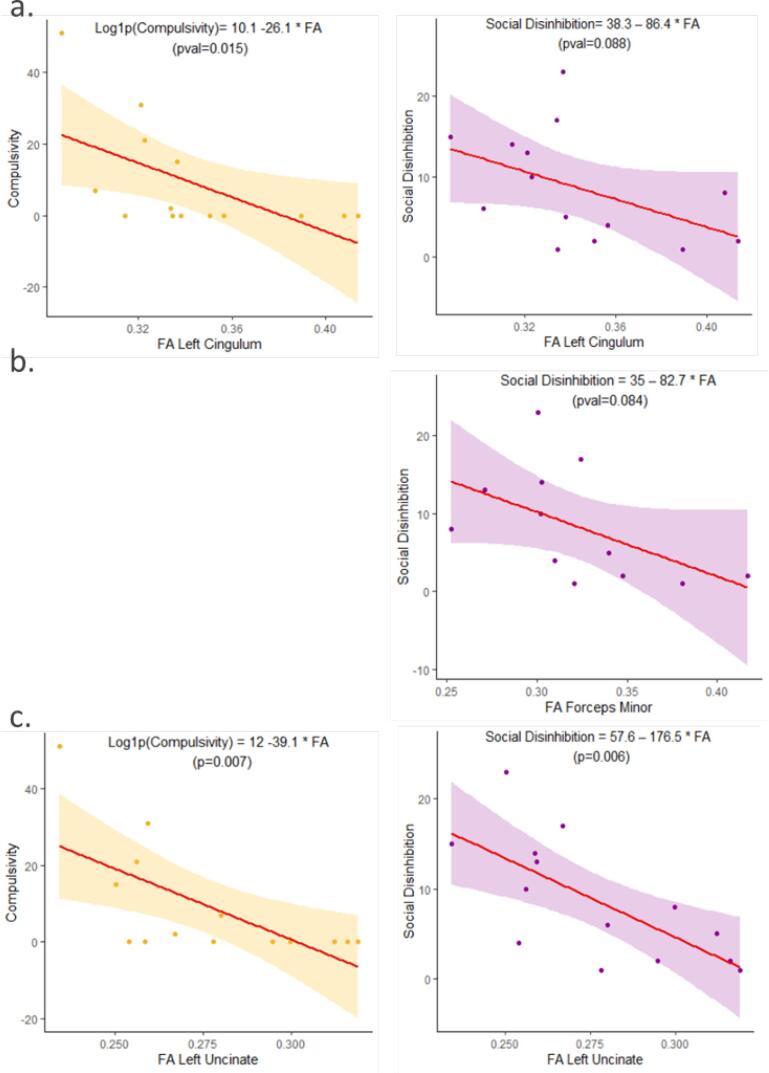
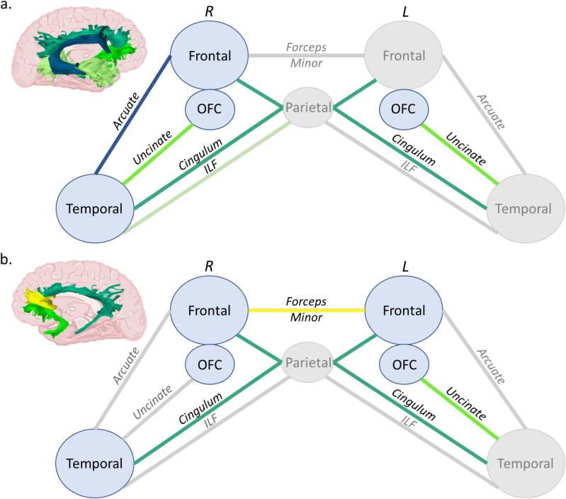
Disinhibition is a core symptom of many neurodegenerative diseases, particularly frontotemporal dementia, and is a major cause of stress for caregivers. While a distinction between behavioural and cognitive disinhibition is common, an operational definition of behavioural disinhibition is still missing. Furthermore, conventional assessment of behavioural disinhibition, based on questionnaires completed by the caregivers, often lacks ecological validity. Therefore, their neuroanatomical correlates are non-univocal. In the present work, we used an original behavioural approach in a semi-ecological situation to assess two specific dimensions of behavioural disinhibition: compulsivity and social disinhibition. First, we investigated disinhibition profile in patients compared to controls. Then, to validate our approach, compulsivity and social disinhibition scores were correlated with classic cognitive tests measuring disinhibition (Hayling Test) and social cognition (mini-Social cognition & Emotional Assessment). Finally, we disentangled the anatomical networks underlying these two subtypes of behavioural disinhibition, taking in account the grey (voxel-based morphometry) and white matter (diffusion tensor imaging tractography). We included 17 behavioural variant frontotemporal dementia patients and 18 healthy controls. We identified patients as more compulsive and socially disinhibited than controls. We found that behavioural metrics in the semi-ecological task were related to cognitive performance: compulsivity correlated with the Hayling test and both compulsivity and social disinhibition were associated with the emotion recognition test. Based on voxel-based morphometry and tractography, compulsivity correlated with atrophy in the bilateral orbitofrontal cortex, the right temporal region and subcortical structures, as well as with alterations of the bilateral cingulum and uncinate fasciculus, the right inferior longitudinal fasciculus and the right arcuate fasciculus. Thus, the network of regions related to compulsivity matched the "semantic appraisal" network. Social disinhibition was associated with bilateral frontal atrophy and impairments in the forceps minor, the bilateral cingulum and the left uncinate fasciculus, regions corresponding to the frontal component of the "salience" network. Summarizing, this study validates our semi-ecological approach, through the identification of two subtypes of behavioural disinhibition, and highlights different neural networks underlying compulsivity and social disinhibition. Taken together, these findings are promising for clinical practice by providing a better characterisation of inhibition disorders, promoting their detection and consequently a more adapted management of patients.
Keywords: Compulsivity; Diffusion tensor imaging; Semi-ecological situation; Social disinhibition; Voxel-based morphometry.
Copyright © 2022 The Authors. Published by Elsevier Inc. All rights reserved.
Conflict of interest statement
The authors declare that they have no known competing financial interests or personal relationships that could have appeared to influence the work reported in this paper.
Figures
References
-
- Miller, B., Llibre Guerra JJ. Frontotemporal dementia. In: Handbook of Clinical Neurology. Vol 165. Elsevier; 2019:33-45. doi:10.1016/B978-0-444-64012-3.00003-4. - PubMed
-
- Rascovsky K., Hodges J.R., Knopman D., Mendez M.F., Kramer J.H., Neuhaus J., van Swieten J.C., Seelaar H., Dopper E.G.P., Onyike C.U., Hillis A.E., Josephs K.A., Boeve B.F., Kertesz A., Seeley W.W., Rankin K.P., Johnson J.K., Gorno-Tempini M.-L., Rosen H., Prioleau-Latham C.E., Lee A., Kipps C.M., Lillo P., Piguet O., Rohrer J.D., Rossor M.N., Warren J.D., Fox N.C., Galasko D., Salmon D.P., Black S.E., Mesulam M., Weintraub S., Dickerson B.C., Diehl-Schmid J., Pasquier F., Deramecourt V., Lebert F., Pijnenburg Y., Chow T.W., Manes F., Grafman J., Cappa S.F., Freedman M., Grossman M., Miller B.L. Sensitivity of revised diagnostic criteria for the behavioural variant of frontotemporal dementia. Brain. 2011;134(9):2456–2477. - PMC - PubMed
Publication types
MeSH terms
LinkOut - more resources
Full Text Sources
Medical
