. 2022 Jun 23;185(13):2279-2291.e17.
doi: 10.1016/j.cell.2022.05.019. Epub 2022 May 27.

Structure, receptor recognition, and antigenicity of the human coronavirus CCoV-HuPn-2018 spike glycoprotein

Affiliations

Structure, receptor recognition, and antigenicity of the human coronavirus CCoV-HuPn-2018 spike glycoprotein

M Alejandra Tortorici et al. Cell. .

Abstract

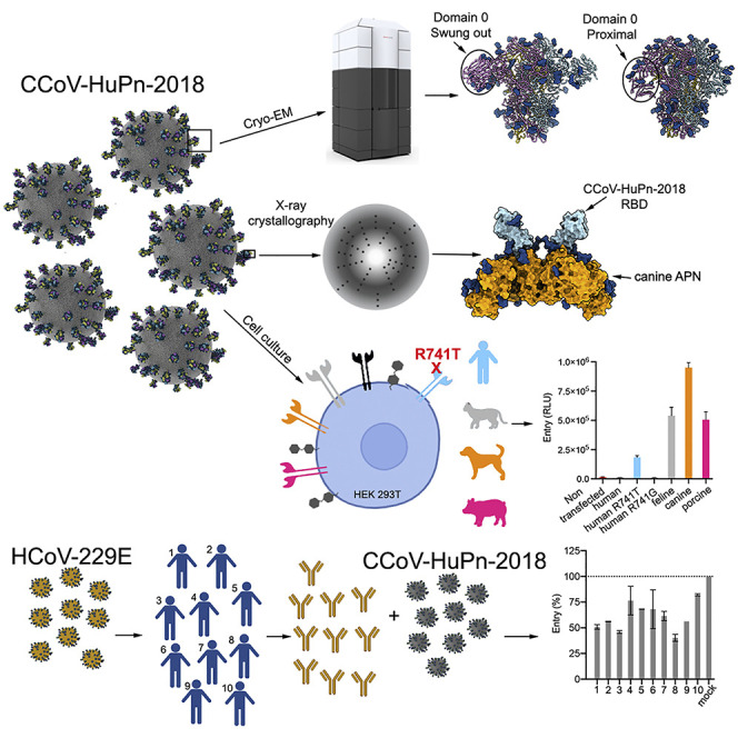
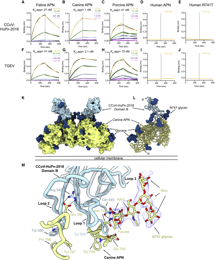
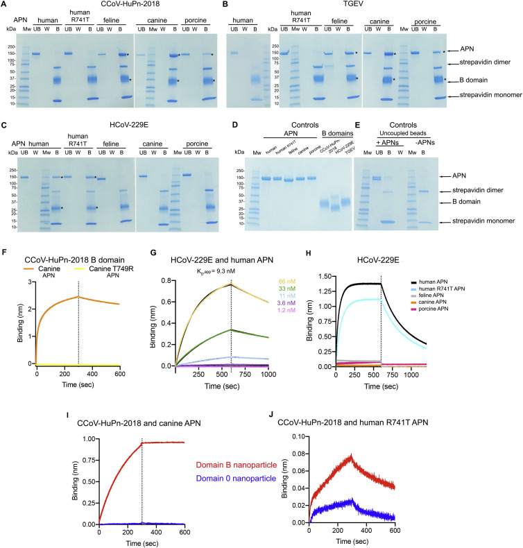
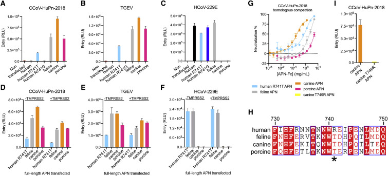
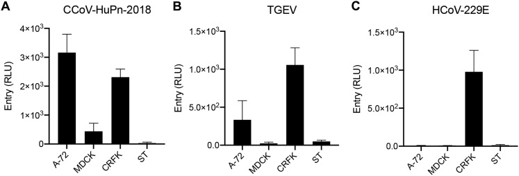
The isolation of CCoV-HuPn-2018 from a child respiratory swab indicates that more coronaviruses are spilling over to humans than previously appreciated. We determined the structures of the CCoV-HuPn-2018 spike glycoprotein trimer in two distinct conformational states and showed that its domain 0 recognizes sialosides. We identified that the CCoV-HuPn-2018 spike binds canine, feline, and porcine aminopeptidase N (APN) orthologs, which serve as entry receptors, and determined the structure of the receptor-binding B domain in complex with canine APN. The introduction of an oligosaccharide at position N739 of human APN renders cells susceptible to CCoV-HuPn-2018 spike-mediated entry, suggesting that single-nucleotide polymorphisms might account for viral detection in some individuals. Human polyclonal plasma antibodies elicited by HCoV-229E infection and a porcine coronavirus monoclonal antibody inhibit CCoV-HuPn-2018 spike-mediated entry, underscoring the cross-neutralizing activity among ɑ-coronaviruses. These data pave the way for vaccine and therapeutic development targeting this zoonotic pathogen representing the eighth human-infecting coronavirus.

Keywords: CCoV-HuPn-2018; HCoV-229E; aminopeptidase; cryo-EM; sialosides; zoonotic viruses; ɑ-coronaviruses.


Conflict of interest statement

Declaration of interests A.T., A.L., and D.C. are employees of Vir Biotechnology Inc. and may hold shares in Vir Biotechnology Inc. D.C. is currently listed as an inventor on multiple patent applications, which disclose the subject matter described in this manuscript. The Veesler laboratory has received a sponsored research agreement from Vir Biotechnology Inc. The remaining authors declare that the research was conducted in the absence of any commercial or financial relationships that could be construed as a potential conflict of interests.

Figures

None
Graphical abstract
Figure 1
Figure 1
Architecture of the CCoV-HuPn-2018 infection machinery (A) Schematic diagram of the S glycoprotein organization. UH, upstream helix; FP, fusion peptide; HR1, heptad-repeat 1; CH, central helix; BH, β-hairpin; CD, connector domain; HR2, heptad-repeat 2; TM, transmembrane domain; CT, cytoplasmic tail. Gray boxes denote regions unresolved in the reconstruction (HR2) and regions that were not part of the construct (TM and CT), respectively. (B and C) Cryo-EM structure of the CCoV-HuPn-2018 S ectodomain trimer (with each domain 0 swung out) viewed along two orthogonal orientations. (D and E) Cryo-EM structure of the CCoV-HuPn-2018 S ectodomain trimer (with each domain 0 pointing “proximal” toward the viral membrane) viewed along two orthogonal orientations. (F and G) Ribbon diagram of the CCoV-HuPn-2018 S1 subunit with domain 0 in the swung out (F) and proximal (G) conformations. (H) Superimposition of the S1 subunit from the structures with domain 0 in the swung out (light blue) and proximal (pink) conformations showing the A domain moving away from the B domain of the same protomer and rotation of domain 0. Renderings in (B–H) use composite models obtained from the global and local refinements for each conformation. (I) Western blot of VSV pseudotyped particles harboring HCoV-229E S, CCoV-HuPn-2018 S, SARS-CoV-2 S, and MERS-CoV S. MW, molecular weight ladder. Full-length S and S2 subunit bands are indicated on the right-hand side of the blot. See also Figures S1 and S2 and Tables S1 and S2.
Figure S1
Figure S1
Data processing and validation of the CCoV-HuPn-2018 S cryo-EM dataset, related to Figures 1 and 2 (A and B) Representative electron micrograph (A) and class averages (B) of CCoV-HuPn-2018 S embedded in vitreous ice. Scale bar of the micrograph, 500 Å. Scale bar of the class averages, 100 Å. (C and D) Gold-standard Fourier shell correlation curves for the CCoV-HuPn-2018 S trimer (solid black line) and the locally refined reconstruction of the CCoV-HuPn-2018 S domain 0 (dashed black line) for the swung out conformation (C) and the proximal conformation (D). The 0.143 cutoff is indicated by a horizontal dashed gray line. (E–H) Local resolution maps for the CCoV-HuPn-2018 S trimer and the locally refined reconstruction of the domain 0 in the swung-out conformation (E and F) and in the proximal conformation (G and H). (I) Cryo-EM data processing flowchart.
Figure S2
Figure S2
Architecture of α-coronavirus S trimers harboring a domain 0, related to Figures 1 and 2 (A–F) Side views of CCoV-HuPn-2018 S with the two conformations of domain 0 (A and D), PEDV S (B, PDB 6VV5), FIPV S (C, PDB 6JX7), PEDV S (E, PDB 6U7K), HCoV-NL63 S (F, PDB 5SZS) perpendicular to the molecular 3-fold axis. Each α-coronavirus S protomer is colored distinctly (light blue, pink, and gold). N-linked glycans are shown as blue spheres. Renderings in (A) and (D) use composite models obtained from the global and local refinements for each conformation.
Figure 2
Figure 2
Structural conservation of CCoV-HuPn-2018 S domain 0 and domain A (A–C) Ribbon diagrams of the CCoV-HuPn-2018 S domain 0 (A) and domain A (B) oriented as shown in the prefusion S conformation in (C). Rendering in (C) uses a composite model obtained from the global and local refinements. (D) Sequence alignment of CCoV-HuPn-2018 S domain A and domain 0 highlighting the low sequence identity (12.5%) between them. (E–G) Topology diagrams of domain 0 (E) and domain A (F) and structural overlay between the two domains (G) underscoring their similarity. N-linked glycans are rendered as blue spheres in (A)–(C) but were removed from (G) for clarity. See also Figures S1 and S2 and Tables S1 and S2.
Figure S3
Figure S3
Architecture of α-coronavirus B domains, related to Figures 1 and 4 (A–C) Ribbon diagrams of the CCoV-HuPn-2018 B domain crystal structure (A), the PRCV B domain bound to porcine APN (PDB: 4F5C) (B), and overlay (C) underscoring their structural similarity. N-linked glycans are rendered as blue spheres. (D–G) Ribbon diagrams of B domains from CCoV-HuPn-2018 (D), HCoV-NL63 (PDB 5SZS) (Walls et al., 2016b) (E), HCoV-229E (PDB 6ATK) (Wong et al., 2017) (F), and PDCoV (PDB 6BFU) (Xiong et al., 2018) (G). All B domains are colored light blue and N-linked glycans are rendered as blue spheres. The receptor-binding loops are indicated.
Figure S4
Figure S4
Characterization of CCoV-HuPn-2018 domain 0-, A-, and B-I53-50 nanoparticles, related to Figure 3 Nanoparticles (NPs) were prepared by incubation of domain 0-I53-50A, domain A-I53-50A, or domain B-I53-50A trimers with pentameric I53-50B at molar ratios of 6 (1-μM domain 0-I53-50A + 5-μM I53-50A):1 1:1 and 1:1, respectively, for 30 min at room temperature. Formation of the domain-0-containing NPs required mixing domain 0-I53-50A and I53-50A, whereas only domain A-I53-50A or domain B-I53-50A were used (besides I53-50B) for assembly of domain-A- or domain-B-containing NPs, respectively. (A–C) Representative electron micrographs of negatively stained CCoV-HuPn-2018 domain 0-, A-, or B-I53-50 NPs. Scale bars, 200 nm. (D) SDS-PAGE analysis of purified nanoparticles.
Figure 3
Figure 3
CCoV-HuPn-2018 S domain 0 recognizes sialic acid and hemagglutinates erythrocytes (A) 50 μL of CCoV-HuPn-2018 domain 0, domain A, or domain B multivalently displayed at the surface of the I53-50 nanoparticle (NP) at 400 ng/μL were incubated with 50 μL of 0.5% (v/v) of turkey, canine, porcine, human, or rat erythrocytes for 30–45 min at room temperature before analysis of hemagglutination. As positive controls, 50 μL of hemagglutinin (HA)-ferritin (HA-ferritin, H1N1 A/New Caledonia/99) and HCoV-HKU1 S NP (HKU-1-S-NP) at 400 and 220 ng/μL were mixed with 50 μL of 0.5% (v/v) turkey or rat erythrocytes, respectively. (B) Serial dilutions of CCoV-HuPn-2018 S domain 0-I53-50 NP (starting at 200 ng/μL) were incubated with the indicated erythrocytes to assess hemagglutination. (C) Serial dilutions of CCoV-HuPn-2018 S domain 0 NP (starting at 100 ng/μL) were incubated with Arthrobacter ureafaciens neuraminidase (NA)-treated human erythrocytes to assess hemagglutination. Erythrocytes mixed with PBS were used as a negative control. Wells positive for hemagglutination are encircled. The experiments were carried out with two biological replicates (using distinct protein and erythrocyte batches) each with two technical replicates. See also Figure S4.
Figure 4
Figure 4
The CCoV-HuPn-2018 S B domain recognizes APN (A–J) Biolayer interferometry kinetic analysis of feline (A and F), canine (B and G), porcine (C and H), human (D and I), and human R741T (E and J) dimeric APN ectodomains binding to biotinylated CCoV-HuPn-2018 B domain (A–E) or TGEV B domain (F–J) immobilized at the surface of SA biosensors. (K–L) Surface representation (K) and ribbon diagram (L) of the crystal structure of CCoV-HuPn-2018 B domain (light blue) in complex with the canine APN ectodomain (light yellow). The observed crystallographic dimer is depicted in its likely orientation relative to the plasma membrane in (K). N-linked glycans are depicted as dark blue surfaces. (M) Close-up view showing key interactions formed between CCoV-HuPn-2018 B domain and the canine APN ectodomain. Dashed lines show salt bridges and hydrogen bonds. The 2Fo-Fc electron density around the N747 glycan is shown as a blue mesh. See also Figures S3 and S5 and Tables S3 and S4.
Figure S5
Figure S5
The CCoV-HuPn-2018 B domain recognizes APN, related to Figures 4 and 5 (A–E) Coomassie-stained SDS-PAGE analysis of pull-downs between biotinylated B domains from CCoV-HuPn-2018 (A), TGEV (B), and HCoV-229E (C) immobilized on magnetic streptavidin beads and APN-Fc orthologs. Asterisks indicate the positions of the pulled down APN/B domain complex. UB, unbound; W, wash; B, bound. Purified proteins used in the pull-down assay (D) and beads without B domain (uncoupled) incubated or not with APNs analyzed by Coomassie-stained SDS-PAGE (E) are also shown. (F) Biolayer interferometry binding analysis of 1-μM wildtype or T749R canineAPN-Fc (glycan knockout) to biotinylated CCoV-HuPn-2018 B domain immobilized at the surface of SA biosensors. (G) Biolayer interferometry kinetic binding analysis of human APN-Fc to biotinylated HCoV-229E B domain immobilized at the surface of SA biosensors. (H) Biolayer interferometry binding analysis of 1 μM human, human R741T, feline, canine, and porcine APN ectodomains (with cleaved Fc fragment) to biotinylated HCoV-229E B domain immobilized at the surface of SA biosensors. (I and J) Biolayer interferometry binding analysis of CCoV-HuPn-2018 domain B- or domain 0-I53-50 nanoparticles to canine APN-Fc (I) or human R741T (J) APN-Fc ectodomains, immobilized at the surface of protein A biosensors.
Figure 5
Figure 5
APN is a functional entry receptor for CCoV-HuPn-2018 (A–C) Entry of VSV particles pseudotyped with CCoV-HuPn-2018 S (A), TGEV S (B), or HCoV-229E S (C) in HEK293T cells transiently transfected with human, human R741T (glycan knockin), human R741G, feline, canine, or porcine APN orthologs. RLUs, relative luciferase units. (D–F) Entry of VSV particles pseudotyped with CCoV-HuPn-2018 S (D), TGEV S (E), and HCoV-V229E S (F) in HEK293T cells transiently transfected with human R741T (glycan knockin), canine, feline, or porcine APN orthologs in the presence or absence of TMPRSS2. (G) Concentration-dependent inhibition of CCoV-HuPn-2018 S pseudovirus entry in HEK293T cells transiently transfected with full-length APN orthologs with matched, purified dimeric soluble APN-Fc ectodomains. (H) Sequence alignment of human, feline, canine, and porcine APNs focused on the N739 glycosylation sequon. Human APN position 741 is indicated with an asterisk. Residue numbering corresponds to human APN. (I) Entry of VSV particles pseudotyped with CCoV-HuPn-2018 S in HEK293T cells transiently transfected with canine or canine T749R glycan knockout mutant (equivalent to human APN position 741). Mean and standard deviation of technical duplicates are graphed. See also Figures S3, S5, and S6 and Tables S3 and S4.
Figure S6
Figure S6
Cell tropism of CCoV-HuPn-2018, TGEV, and HCoV-229E S pseudoviruses, related to Figure 5 (A–C) Evaluation of pseudotyped virus mediated entry in feline, canine, and porcine cell lines. CCoV-HuPn-2018 S (A), TGEV S (B), and HCoV-229E S (C) VSV pseudotyped virus entry in Canis familiaris tumor fibroblast cells (A-72), Canis familiaris Madin-Darby canine kidney (MDCK), and Felis catus Crandell-Rees feline kidney (CRFK) epithelial kidney cells and Sus scrofa pig testis fibroblast cells (ST). Mean and standard deviation of technical duplicates are graphed.
Figure 6
Figure 6
Evaluation of polyclonal and monoclonal antibody neutralization of CCoV-HuPn-2018 S-mediated entry into cells (A and B) Sequence conservation of HCoV-229E, HCoV-NL63, and CCoV-HuPn-2018 S glycoproteins plotted on the CCoV-HuPn-2018 S structure viewed from the side (A) and top (B). The sequence alignment was generated using the following sequences: CCoV-HuPn-2018 S (QVL91811.1), HCoV-229E S (AAK32191.1 and ABB90515), and HCoV-NL63 S (AIW52835.1 and YP_003767.1). Renderings in (A) and (B) use a composite model obtained from the global and local refinements. (C–E) HCoV-229E S (C), CCoV-HuPn-2018 S (D), and TGEV S (E) pseudotyped VSV entry in the presence of a 1:5-diluted plasma obtained between 1985 and 1987 from human subjects previously infected with HCoV-229E (dose-response neutralization curves are shown in Figure S7). (F and G) CCoV-HuPn-2018 S (F) and TGEV S (G) pseudotyped VSV entry in the presence of various concentrations of 1AF10 neutralizing monoclonal Fab fragment in HEK293T cells transfected with the indicated full-length APN orthologs. (H and I) Competition between 1AF10 Fab and APN for binding to immobilized CoV-HuPn-2018 (H) or TGEV (I) B domains analyzed by biolayer interferometry. Each CoV-HuPn-2018 (H) or TGEV (I) B-domain-loaded SA biosensor was sequentially dipped in a solution containing the 1AF10 Fab at a concentration ten times above the respective affinity (170 nM for CoV-HuPn-2018 or 100 nM for TGEV), and then a solution containing the same concentration of 1AF10 supplemented with canine APN (orange, 10 nM for CoV-HuPn-2018 or 21 nM for TGEV), feline APN (gray, 270 nM for CoV-HuPn-2018 or 470nM for TGEV), porcine APN (pink, 410 nM for CoV-HuPn-2018 or 700 nM for TGEV), or no APN (blue). APNs used here had the Fc tag cleaved. Mean and standard deviation of technical duplicates are graphed. See also Figure S7.
Figure S7
Figure S7
Inhibition of CCoV-HuPn-2018-S-mediated entry into cells by human polyclonal plasma antibodies and binding of CCoV-HuPn-2018 B domain to a monoclonal antibody, related to Figure 6 (A–C) CCoV-HuPn-2018 (A), TGEV (B), and HCoV-229E (C) S VSV pseudotyped mediated entry in the presence of various dilutions of plasma (name of the sample is indicated at the top of each graph) obtained between 1985 and 1989 from human subjects previously infected with HCoV-229E. One representative experiment out of two biological replicates is shown. Fits are shown only when inhibition of entry was observed. (D and E) Biolayer interferometry kinetic analysis of 1AF10 Fab binding at various concentrations to biotinylated CCoV-HuPn-2018 (D) or TGEV (E) B domains immobilized at the surface of SA biosensors.
Figure S8
Figure S8
Glycan-mediated immune evasion strategy of α-coronaviruses, related to Figure 1 (A–C) Ribbon diagrams of S1 subunits from HCoV-NL63 S (PDB 5SZS) (A) and from CCoV-HuPn-2018 S with domain 0 in proximal (B) or swung out (C) conformations. The HCoV-NL63 receptor-binding loops are buried through interactions with domain A of the same protomer (light blue), including the glycan moiety at Asn358, and are not available to engage host cell receptors. The CCoV-HuPn-2018 receptor-binding loops are also buried by interactions with domain A, including the glycan at position N404 of the same protomer (light blue) and N561 from another protomer (pink). Renderings in (B) and (C) use composite models obtained from the global and local refinements for each conformation.

References

    1. Agirre J., Iglesias-Fernández J., Rovira C., Davies G.J., Wilson K.S., Cowtan K.D. Privateer: software for the conformational validation of carbohydrate structures. Nat. Struct. Mol. Biol. 2015;22:833–834. - PubMed
    1. Anthony S.J., Gilardi K., Menachery V.D., Goldstein T., Ssebide B., Mbabazi R., Navarrete-Macias I., Liang E., Wells H., Hicks A., et al. Further evidence for bats as the evolutionary source of Middle East respiratory syndrome coronavirus. mBio. 2017;8 e00373–e00317. - PMC - PubMed
    1. Armon A., Graur D., Ben-Tal N. ConSurf: an algorithmic tool for the identification of functional regions in proteins by surface mapping of phylogenetic information. Journal of Molecular Biology. 2001;307:447–463. - PubMed
    1. Arunachalam P.S., Walls A.C., Golden N., Atyeo C., Fischinger S., Li C., Aye P., Navarro M.J., Lai L., Edara V.V., et al. Adjuvanting a subunit COVID-19 vaccine to induce protective immunity. Nature. 2021;594:253–258. - PubMed
    1. Bale J.B., Gonen S., Liu Y., Sheffler W., Ellis D., Thomas C., Cascio D., Yeates T.O., Gonen T., King N.P., et al. Accurate design of megadalton-scale two-component icosahedral protein complexes. Science. 2016;353:389–394. - PMC - PubMed

Publication types