

A Rare Cause of Chest Pain Identified on Point-of-care Echocardiography: A Case Report
- PMID: 35701360
- PMCID: PMC9197749
- DOI: 10.5811/cpcem.2021.9.53553
A Rare Cause of Chest Pain Identified on Point-of-care Echocardiography: A Case Report
Abstract
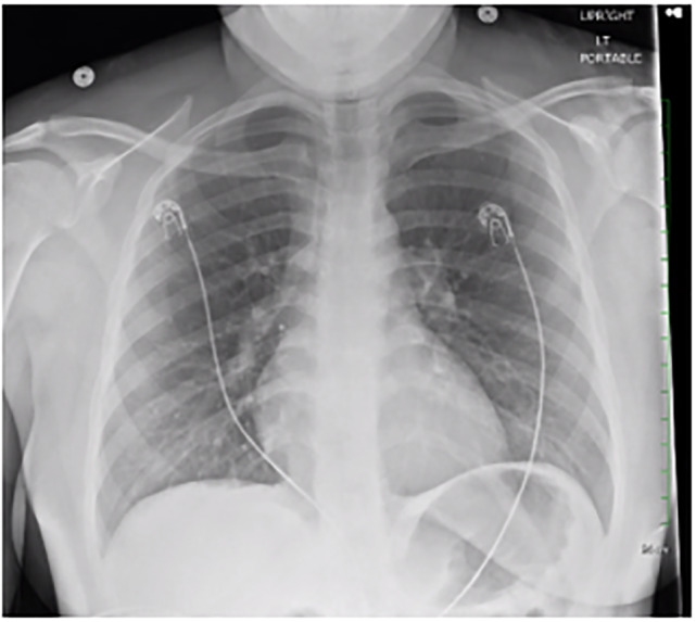
Introduction: Cardiac masses are a rare cause of chest pain. They can often be missed on a chest radiograph performed to evaluate non-specific chest pain and are not readily evaluated with traditional laboratory testing. However, these masses can be visualized with point-of-care ultrasound.
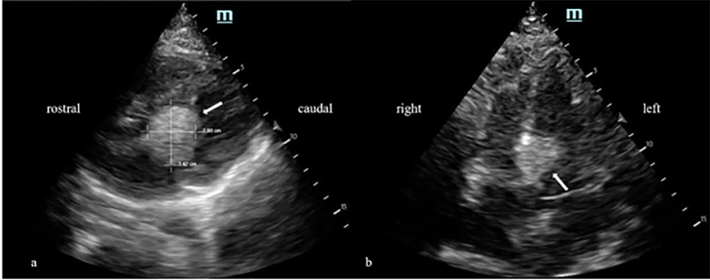
Case report: We present a case of a 19-year-old female presenting with intermittent chest pain, palpitations, and weakness present for two months. The patient had previously been evaluated at our emergency department one week earlier and was diagnosed with anxiety before being discharged. Besides a tachycardic and labile heart rate, physical examination and laboratory testing were unremarkable. Point-of-care cardiac echocardiography subsequently demonstrated findings concerning for a cardiac mass.
Conclusion: Cardiac masses are a rare cause of chest pain and palpitations that are easily missed. The advent of point-of-care ultrasonography has afforded us the ability to rapidly assess for structural and functional cardiac abnormalities at bedside, and incorporation of this tool into the evaluation of patients with chest pain offers the ability to detect these rare pathologies.
Conflict of interest statement
Figures
Similar articles
-
Hemorrhagic Pericardial Cyst Diagnosis Accelerated by Emergency Physician Echocardiography: A Case Report.J Emerg Med. 2017 Apr;52(4):e105-e109. doi: 10.1016/j.jemermed.2016.10.023. Epub 2017 Jan 20. J Emerg Med. 2017. PMID: 28117110
-
Assessing sensitivity and specificity of the Manchester Triage System in the evaluation of acute coronary syndrome in adult patients in emergency care: a systematic review protocol.JBI Database System Rev Implement Rep. 2015 Nov;13(11):64-73. doi: 10.11124/jbisrir-2015-2213. JBI Database System Rev Implement Rep. 2015. PMID: 26657465
-
Evaluation of chest pain in the emergency department.Curr Probl Cardiol. 1997 Apr;22(4):149-236. doi: 10.1016/s0146-2806(97)80007-2. Curr Probl Cardiol. 1997. PMID: 9107535 Review.
-
Inferior Vena Cava Filter Limb Fracture with Embolization to the Right Ventricle.J Emerg Med. 2017 Aug;53(2):248-251. doi: 10.1016/j.jemermed.2017.01.047. Epub 2017 Mar 6. J Emerg Med. 2017. PMID: 28279544
-
[Is a more efficient operative strategy feasible for the emergency management of the patient with acute chest pain?].Ital Heart J Suppl. 2000 Feb;1(2):186-201. Ital Heart J Suppl. 2000. PMID: 10731376 Review. Italian.